The great majority of sport-related ruptures of the anterior cruciate ligament (ACL) occur when the injured knee has not been struck directly by an opponent or by equipment. ACL rupture without a direct blow to the knee cannot be explained by an external force in a single plane acting alone. The core mechanism is that, during a cutting stop or a single-leg landing, axial compression in shallow flexion, anterior pull by the quadriceps, a knee-abduction (valgus) moment, and a tibial internal-rotation moment convert into one another through the anatomy and tighten the ACL together; in cadaveric experiments with repeated high-load impacts, accumulation of microdamage at the bony attachments or in the fibers can lower the tissue's tolerance limit, a process that is compatible with acute multiplanar overload and that jointly contributes to structural failure[3,9–13,16,17]. Visible limb alignment is not the true intra-articular moment, and neither a local peak strain measured by a cadaveric sensor nor a late joint-collapse frame on match video can be equated directly with the precise instant of ligament tear in vivo.
1. Evolution of Contact Classification: From "No Direct Lower-Limb Contact" to a Three-Category System
How the literature defines a "noncontact" injury has changed in substance over the past twenty-odd years, and that change directly affects comparison of percentage figures across studies. Early video studies in handball and basketball mostly used "no direct impact to the injured lower limb or knee" as the dividing rule: Olsen et al.[1], analyzing 20 injury videos from women's handball and interviews with 32 players, assigned a shove to the trunk or a pull on an upper limb to local disturbance; Krosshaug et al.[2], analyzing 39 basketball videos (22 women, 17 men), separately counted body collisions and pushes before ground contact (11/22 women, 1/17 men) and estimated body contact at the injury frame (6 women, 5 men, including 4 male players with a direct blow to the injured knee). Koga et al.[3], in a model-based image-matching (MBIM) study of 10 women's handball and basketball cases, retained the older definition of "no direct lower-limb contact" and labeled all 10 noncontact; yet Table 1 of that paper (PDF p. 4) shows that in cases 3, 4, 5, 7, 8 and 10—six players in all—there was a shove to the trunk, a body collision or a pull on the jersey before or at ground contact, and only 4 cases had no body contact at all.
To distinguish the disturbance that body contact produces in whole-body motor control, Waldén et al.[4] (39 men's soccer videos), Della Villa et al.[5] (134 men's soccer videos) and Sundberg et al.[6] (a systematic review covering 20 sports, 62 studies and 5612 injury situations) adopted a three-category standard:
- Noncontact injury: apart from the normal contact of the sole with the ground or a mat, there is no person-to-person or person-to-object contact anywhere on the body during the injury event;
- Indirect-contact injury: the injured knee is not struck directly by an external force, but before ground contact or at the instant of injury the trunk, pelvis, upper limb, contralateral lower limb, or an ipsilateral segment other than the injured knee is hit, pushed or pulled;
- Direct-contact injury: an external force acts directly on the injured knee (for direct-impact mechanisms, see Section 1.10).
Statistics in contact sports are also affected by the time window used to judge contact. Waldén et al.[4], recording status at the estimated injury frame (IF), reported 25 noncontact (64%), 8 indirect-contact (21%) and 6 direct-contact (15%) cases. Della Villa et al.[5], among 134 cases, finally classified 59 as noncontact (44.0%), 59 as indirect contact (44.0%) and 16 as direct contact (11.9%); if classified only at the estimated injury frame, there were 87 with no contact and 31 with indirect contact, and the final equal split of 59 each arose because 28 players had already been pushed on the upper limb or trunk during the run-up or flight before the injury frame, with that contact continuing through single-leg landing. The external force in indirect contact does not itself snap the ligament; rather, by disrupting trunk control and landing preparation, it induces multidirectional endogenous loading of the lower limb while the injured knee remains free of a direct blow.
2. Weighing Evidence for Multidirectional Loading Versus Unidirectional Explanations
Whether ACL rupture without a direct blow to the injured knee is driven chiefly by "sagittal-plane axial compression and anterior shear," or by "combined multiplanar loading (anterior shear, a knee-abduction moment and a tibial internal-rotation moment acting together)," is a point of disagreement in the literature. Taking cadaveric biomechanics and in vivo kinematics together, a single plane or a single force vector is hard to reconcile with real injury; axial compression in shallow flexion, anterior pull by the quadriceps, a knee-abduction moment and a tibial internal-rotation moment that convert into one another through the anatomy and tighten the ACL together form the more reliable mechanical account (for the basic tension properties, see Section 1.8).
1. Common Combinations in Shallow Flexion, Sagittal Shear, and Transmission of Axial Compression
In tasks such as a decelerating cut, a single-leg landing after a jump shot, or recovery of balance on the support leg after a kick, the injured knee at initial contact (IC) is often in 0°–30° of shallow flexion (a common high-risk posture, but not a necessary condition for every noncontact injury)[1–6]. In the 0°–30° shallow-flexion range the pull of the patellar tendon is directed markedly anterior to the long axis of the tibia; a strong eccentric quadriceps contraction therefore produces not only axial compression but also a large anterior tibial shear component, while the small attachment angle of the hamstring tendons yields only a small posterior tibial shear component[9].
Boden and Sheehan[10] and Beaulieu et al.[9] further set out how axial compression is converted, through the posteriorly sloping tibial plateau, into anterior shear and rotation: the lateral tibial plateau slopes posteriorly and inferiorly and its articular surface is slightly convex, whereas the anterior part of the lateral femoral condyle is relatively flat; under high-magnitude axial compression in shallow flexion the contact point of the lateral femoral condyle moves forward and the condyle slides posteriorly and inferiorly along the sloping lateral plateau (the tibia advances relative to the femur); the medial plateau is concave and more constrained, so preferential anterior translation of the lateral compartment produces concomitant tibial internal rotation and tightens the ACL[9,10]. When an athlete, because of a sudden stop or a disturbance, shifts the trunk posteriorly and increases hip flexion, the moment arms of the ground-reaction force relative to the hip and knee change accordingly. In the case–control videos summarized by Boden et al.[10], injured players often landed on a flat foot or on the heel (mean plantar-flexion angle of the ankle at contact 11°, controls 23°, P = 0.0059), shortening the time available for eccentric buffering by the triceps surae and transmitting ground impact more directly to the knee; flat-foot or heel contact is not, however, a universal premise—Waldén et al.[4], in men's soccer videos, observed that injury on a single-leg landing after a contested header could begin with forefoot contact, followed rapidly in stance by ankle dorsiflexion and valgus instability of the knee.
Defects of argument and mixed comparison of data in Boden and Sheehan's 2022 review[10] (PDF pp. 4, 5, 7, 8). That paper has three defects that affect its conclusions: (1) mixed comparison across variables and designs (PDF pp. 5, 8)—the authors divided the 30° difference in dynamic spatial inclination of the tibial plateau relative to the vertical under gravity between simulated "high-risk" and "safe" postures on standing MRI in 25 healthy athletes by the 1.5° between-group mean difference in intrinsic bony posterior tibial slope reported in the literature between injured and healthy groups (30 ÷ 1.5) and called the dynamic postural effect "20 times" the bony slope; the two quantities belong to different physical variables and study designs, and direct division is not valid; (2) equating cadaveric uniaxial failure force directly with external ground-reaction force (PDF p. 5)—they converted a cadaveric femur–ACL–tibia uniaxial failure load (2160 N) directly into 3.1 body weights of ground-reaction force for a 70 kg athlete, ignoring load sharing by the articular surfaces, menisci, muscle groups and the remaining ligaments; (3) treating a retrospective classification rate as prospective prediction and citing unpublished conclusions (PDF pp. 4, 7, 8)—they called the retrospective discrimination rate for center-of-mass–to–base-of-support distance in 20 injured and 20 control videos an "80% accuracy for predicting injury occurrence," and, on the strength of an unpublished military-academy cohort (reference 41 noted "In Preparation"), asserted that knee extension and valgus are not risk factors. This chapter does not adopt their "20-fold effect," the "2160 N or 3.1 body-weight injury threshold," the "80% prospective prediction rate," or the rejection of multiplanar loading; it retains the qualitative mechanisms that flat-foot landing reduces calf buffering and that the sloping lateral plateau converts axial compression into posterior femoral sliding.
2. Coronal Abduction, Transverse Internal Rotation, and Multiplanar Combined-Loading Experiments
Kiapour et al. 2016[11] (PDF p. 3) and Quatman et al. 2014[12] (PDF p. 3) used the same set of 17 fresh-frozen cadaveric lower limbs (45 ± 7 years; 9 women, 8 men; flexion fixed at 25°; static pretension of 1200 N in the quadriceps and 800 N in the hamstrings) to report ACL strain and the ACL/medial collateral ligament (MCL) strain ratio, respectively, and cannot be added to make 34 independent specimens; Kiapour et al. 2014[13] tested 16 specimens on the same apparatus (45 ± 7 years; 8 women, 8 men); the finite-element model of Quatman et al.[12] was built from the anatomy of one healthy 25-year-old female athlete.
Kiapour et al. 2016[11] implanted a differential variable reluctance transducer (DVRT) in the anteromedial (AM) bundle of the ACL and simulated bilateral landing axial impact with a 350 N weight dropped from 30 cm (peak about 3–4.2 kN). Under the baseline condition of muscle pretension and axial impact alone, local peak ACL strain was 6.8% ± 1.0% (mean ± standard error of the mean, SEM); when an isolated 268 N anterior tibial shear force, a 75 N·m knee-abduction moment or a 60 N·m tibial internal-rotation moment was added, peak strain rose to 10.6% ± 1.1% (P = 0.009), 12.4% ± 1.7% (P = 0.009) and 10.9% ± 2.9% (P = 0.049), respectively; when all three were applied together (268 N anterior shear + 75 N·m abduction + 60 N·m internal rotation), peak strain rose to 16.9% ± 1.7%, a mean higher than each of the three unidirectional loading levels (Figure 1); between-group comparison pooled by loading mode likewise showed that the multiplanar combined-loading group was significantly higher than each unidirectional group (P < 0.001)[11].
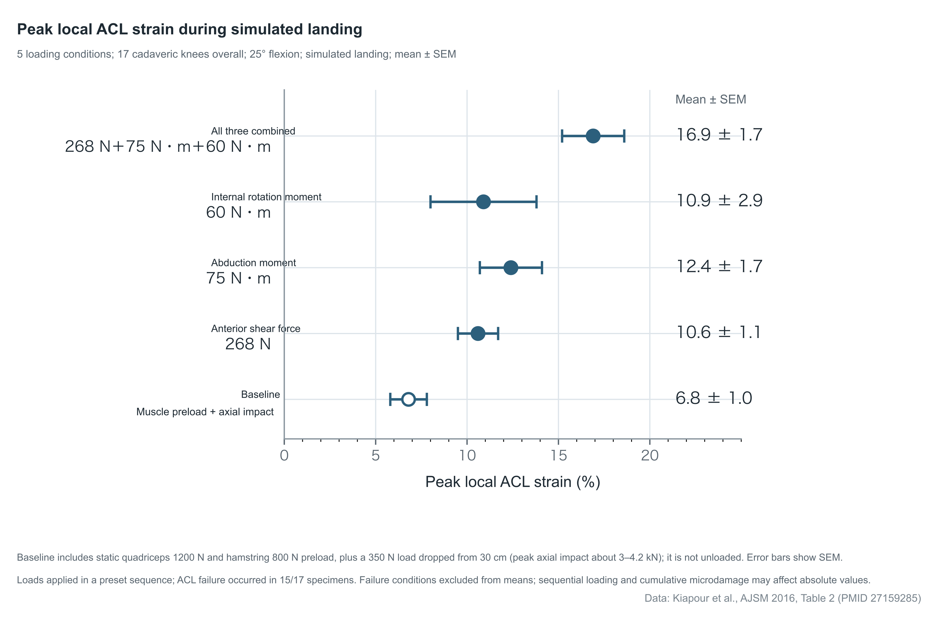
Figure 1. Comparison of local peak strain in the anteromedial bundle of the ACL under uniplanar and multiplanar loading during simulated cadaveric landing. Data from Table 2 (PDF p. 6) of Kiapour et al. 2016[11]; whole cohort of 17 human cadaveric lower limbs (25° of flexion), each with 1200 N quadriceps and 800 N hamstring pretension and a 350 N drop from 30 cm producing axial impact (baseline load nonzero). Dots and error bars are mean ± SEM; ACL failure occurred in 15/17 specimens, and failure conditions were excluded from the means (effective sample size under each condition is therefore not uniformly 17). The values represent local strain under this specific experimental configuration only and are not a clinical rupture threshold.
Methodological limits and typographical errors in the data of Kiapour et al. 2016[11] (PDF Table 2 p. 6, and pp. 7 and 9). (1) Repeated addition of the baseline and absence of a formal interaction test (PDF p. 9)—the authors added the three uniplanar total peak strains directly (10.6% + 12.4% + 10.9% = 33.9%, written in the text as "about 34%") and contrasted the sum with combined loading (16.9%), but each uniplanar total strain already includes the 6.8% baseline strain, so direct addition counts the baseline three times; the paper also built no interaction-term model for an interaction test. (2) Coefficient of determination and typographical errors in Table 2 (PDF Table 2 p. 6 and p. 7)—on p. 7 the text misprints the coefficient of determination of internal-rotation angle for ACL strain, R² = 0.32 (see the Abstract and Fig. 4B, explaining 32% of the variance), as 0.32%; in Table 2 on p. 6, the 95% confidence interval for ACL strain 14.5 ± 1.8 in the 0 N / 75 N·m / 20 N·m row is printed as (0.8–18.2), which is inconsistent with the mean and SEM; the 95% confidence interval for internal-rotation angle 34.9 ± 2.3 in the 268 N / 125 N·m / 60 N·m row is misprinted as (30.3–30.5) (upper bound below the mean); the standard error in the 0 N / 75 N·m / 60 N·m row is printed as 25.9 ± 2,6; and the 268 N / 0 N·m / 20 N·m row is printed as 17.3-3.0. (3) Scope of interpretation—abduction and internal rotation were static pretension applied before impact, and the univariate regression slopes (abduction angle β = 0.42 versus internal-rotation angle β = 0.23) are influenced by the pretension levels. This chapter does not adopt the "additive ≈34%" argument, the misprinted intervals in Table 2, the "0.32%" in the text, or an absolute ranking of primacy between abduction and internal rotation; it retains the experimental result that multiplanar combined loading raises local AM-bundle ACL strain significantly more than unidirectional loading.
Addressing why complete MCL tear is uncommon clinically if an abduction moment participates in injury, Quatman et al.[12] measured local strain in the AM bundle of the ACL and in the superficial MCL simultaneously on the same 17 specimens: under every uniplanar and multiplanar loading condition the ACL-to-MCL peak-strain ratio exceeded 1.7, and the relative increase in ACL strain from baseline was significantly greater than that of the MCL (P < 0.01); ACL failure occurred in 15 of 17 specimens (88%) (7 [47%] under three-plane combined loading), and no specimen sustained a complete MCL tear. A companion single-subject finite-element parametric analysis showed that, under 268 N anterior shear and muscle pretension, as the abduction moment or the internal-rotation moment was varied separately, lateral-compartment compression and concomitant internal rotation preferentially tightened the ACL; only in the parametric intervals in which the abduction moment exceeded 48 N·m or the internal-rotation moment exceeded 24 N·m did the slope of MCL strain growth with load begin to exceed that of the ACL (an observation of parametric slopes in a single finite-element model, not a human rupture threshold)[12]. The clinical scarcity of complete MCL rupture is therefore not grounds for excluding a knee-abduction moment from participation in ACL injury.
In millisecond-scale timing, Kiapour et al. 2014[13], measuring 16 cadaveric lower limbs (25° of flexion; pretension and impact as above; no specimen ruptured), showed that axial impact force peaked first at 13.7 ± 2.4 ms after contact (4109 ± 691 N), followed in turn by the increments in knee flexion (36.6 ± 7.8 ms), anterior tibial translation (37.5 ± 7.0 ms), knee abduction (51.5 ± 22.6 ms) and local ACL strain (54.2 ± 27.0 ms), with peak tibial internal rotation at 86.5 ± 25.1 ms (P < 0.015). Although peak internal rotation came later, the text on PDF pp. 2 and 6 of that paper shows that when ACL strain peaked at 54.2 ms, tibial internal rotation had already begun (21.0 ± 11.7 ms) and had reached 1.8° ± 2.5°, about 63% of its final peak (2.8° ± 2.6°)[13]; at the same time, the 54.2 ms strain-peak time in this subfailure experiment cannot be equated directly with the instant of ligament tear in real injury.
3. Temporal Sequence on Injury Video, Visible Valgus, Bone Bruises, and Post-Injury Reduction
Cadaveric models can measure local strain directly but cannot fully reproduce whole-body neuromuscular control on the field; match video and post-injury MRI record real injury events but are limited by sampling frame rate, two-dimensional projection and the phase of observation. Injurious loading early after ground contact and secondary displacement after ligament rupture occur in different phases; how the two appear on video and imaging, and the research basis for each, are compared in Table 1.
Table 1. Comparison of observation of ACL injury–related time points and joint positions across study designs
| Source study and method | Sample and subjects | Core time point or joint position | Nature and limits of interpretation |
|---|---|---|---|
| Koga et al. 2010[3] (MBIM three-dimensional matching) | 10 women's handball/basketball videos (6 with indirect body contact) | From IC to 40 ms, flexion, valgus and internal rotation increased rapidly; after 40 ms the rotation reversed to external; estimated peak ground-reaction force at 40 ms | ≈40 ms is a kinematic inference based on an angular inflection and estimated reaction force, not a visualized tear; early millimeter-scale anterior translation cannot be quantified; case 10 showed no early internal rotation |
| Kiapour et al. 2014[13] (cadaveric drop-weight landing) | 16 cadaveric lower limbs, neutral landing at 25° (no ligament rupture in any) | Axial force 13.7 ms, anterior translation 37.5 ms, abduction 51.5 ms, ACL strain 54.2 ms, internal rotation 86.5 ms peaked in that order | 54.2 ms is a subfailure local strain peak, not equivalent to the clinical rupture instant; at the strain peak, internal rotation had already completed about 63% of its peak amplitude |
| Della Villa et al. 2020[5] (two-dimensional video assessment) | 134 men's soccer videos (107 with biomechanical assessment) | At the preset estimated injury frame (IC+40 ms), 81% showed visible valgus alignment; only 13% showed marked valgus collapse | IC+40 ms is a uniform analysis frame adopted from Koga's inference and does not prove that every case ruptured at 40 ms; visible valgus alignment is not a knee-abduction moment |
| Grassi et al. 2020[14] (two-dimensional video reduction) | 21 cases screened for visible rapid reduction from 137 men's soccer videos in the same parent cohort | Rapid posterior reduction began at 229 ± 81 ms after IC and 55 ± 30 ms after the injured foot left the ground and unloaded, lasting 42 ± 11 ms | Description of a screened visible subset; cannot be taken as an overall incidence or as the tear instant; association of unloading reduction with meniscal injury and a pop is theoretical inference |
| Grassi et al. 2021[15] (MRI bone-bruise matching) | 7 of 62 men's soccer injuries with both video and bicompartmental bone-bruise MRI | Three-dimensional matched bone-bruise position: 46.1° flexion, 4.1° valgus, 6.1° tibial external rotation (control squat: 7.6° internal rotation) | Reflects a bone-surface proximity position estimated in three-dimensional geometry, with no millisecond timestamp; does not prove that the bruise necessarily formed late after rupture; the 46.9 mm coordinate is not a clinical anterior-laxity measure |
1. "Valgus Alignment" and "Valgus Collapse" on Three-Dimensional Image Matching and Two-Dimensional Video
Using multi-camera footage to build three-dimensional skeletal models (MBIM), Koga et al.[3] depicted the injury process in 10 female handball and basketball athletes: at IC mean knee flexion was 23° (11°–30°), the coronal plane was near neutral (0°, −2° to 3°), and tibial external rotation was 5° (−5° to 12°); in the first 40 ms after contact, flexion increased by 24° (95% CI 19°–29°), abduction (valgus) by 12° (95% CI 10°–13°) and tibial internal rotation by 8° (95% CI 2°–14°); after 40 ms there was an abrupt shift to a mean 17° (95% CI 13°–22°) of tibial external rotation and a larger valgus displacement. Combining a vertical ground-reaction force peak estimated from center-of-mass acceleration (3.2 body weights at 40 ms after IC), the authors inferred that ACL rupture occurred within about 40 ms after ground contact[3]. The study is limited by four methodological conditions: vertical ground-reaction force was estimated from the derivative of the center of mass, not measured on a force plate; broadcast-video resolution cannot reliably quantify early millimeter-scale anterior tibial translation; in Fig. 3, case 10 showed no tibial internal rotation in the first 40 ms (Table 2 uses a minus sign for abduction and external-rotation directions, whereas the text reports absolute values—a sign convention); and "rupture at about 40 ms" is an inference from an angular inflection, not direct visualization of ligament tear.
Two-dimensional match-video analysis must distinguish visible coronal valgus alignment from large-amplitude valgus collapse. Della Villa et al.[5] observed coronal knee-valgus alignment at the estimated injury frame (IC + 40 ms) in 81% of assessable men's soccer cases, but large-amplitude valgus collapse in only 13%; Waldén et al.[4] likewise noted that marked valgus collapse was uncommon in 39 men's soccer videos, whereas Krosshaug et al.[2], in a basketball kinematic subsample, recorded obvious valgus collapse in 9/17 women and 2/13 men. Two-dimensional valgus alignment is influenced by camera projection, hip internal rotation and knee flexion angle, and is not a knee-abduction moment; as for the absence of large-amplitude valgus collapse in most male players, one possible clinical explanation is that after early rupture at ground contact the player immediately unloads and falls, so that lack of late large collapse cannot be used to deny participation of early abduction and rotational loading.
2. Bicompartmental Bone-Bruise Matching and Rapid Posterior Tibial Reduction After Foot Lift
Grassi et al. 2021[15] screened 7 patients who had both multi-view video and bicompartmental bone-bruise MRI from 62 men's soccer ACL injuries, reconstructed with a three-dimensional surface-distance minimization algorithm the joint position at which the femoral and tibial bone-bruise centers were closest, and compared it with dynamic biplanar radiostereometric analysis (RSA) of single-leg squatting in ACL-deficient patients. The matched bone-contact position was 46.1° ± 3.8° of flexion, 4.1° ± 2.4° of abduction and 6.1° ± 3.7° of tibial external rotation (control squat at the same flexion angle: 7.6° ± 5.0° of internal rotation, P = 0.04). This deeper-flexion, externally rotated posture is consistent in direction with the secondary displacement after 40 ms post-IC described by Koga et al.[3], suggesting that bicompartmental bone bruises may correspond to a later bone-surface impact state. Three-dimensional bone-bruise matching cannot, however, prove that the bruise necessarily formed late after ligament rupture: the algorithm must assume that the medial and lateral bruises form synchronously in the same instant and lacks cartilage/meniscal deformation data, and MRI itself carries no millisecond-scale temporal information; the anatomic-coordinate origin offset reported in the paper (46.9 ± 2.8 mm) is a modeling coordinate distance and must not be misread as clinical anterior tibial laxity.
Grassi et al. 2020[14], from 137 men's soccer videos in the same parent cohort as Della Villa et al.[5], screened 21 cases by "a rapid posterior tibial jump-reduction recognizable by eye" (16 noncontact, 5 with pre-injury indirect contact; a feature-subset analysis, so 21/137 cannot be used to estimate an overall reduction rate). In the 21 cases, visible rapid posterior tibial reduction began on average 229 ± 81 ms after IC and 55 ± 30 ms after the injured foot left the ground (49 ± 10 ms after excluding one outlier), lasted 42 ± 11 ms, and occurred in flexion beyond 45° in 86%[14]. This shows that the abrupt tibial movement around 200 ms after ground contact is post-injury reduction, not the instant of tear. As for the discussion the authors advanced from prior single-case reports—that early compression and anterolateral subluxation produce ACL rupture, bone bruise and meniscal involvement, and that unloading when the injured foot leaves the ground allows the anteriorly locked tibia to reduce rapidly posteriorly, possibly with a subjective "pop"—the original paper already states that this is theoretical inference, not confirmed by direct mechanical measurement[14].
4. Single Supraphysiologic Overload and Accumulation of Repeated Subfailure Microdamage
Noncontact ACL rupture almost always presents with sudden symptoms during a single cut or landing, but sudden symptoms alone cannot prove that the ligament had no structural degeneration before the last ground contact: overload from a single combined loading event and prior accumulation of microdamage from high-frequency subfailure impacts that lowers tissue threshold must be examined together with cadaveric fatigue experiments and human data.
1. Cadaveric Repeated Pivot Landings and Experiments Restricting Femoral Internal Rotation
A University of Michigan group has shown that the human ACL can fail in low-cycle fatigue under repeated multiplanar loading below the single-cycle failure limit[9,16,17]. Lipps et al. 2013[16] took 10 pairs (20 knees; 5 male pairs, 5 female pairs) of matched cadaveric knees and, at an initial 20° of flexion with cross-knee muscle pretension, applied repeated pivot impacts targeting 3 body weights (3×BW; measured 3.5 ± 0.3 BW with 29.8 ± 5.4 N·m of internal-rotation moment) to one side and 4 body weights (4×BW; measured 4.3 ± 0.4 BW with 34.5 ± 7.3 N·m of internal-rotation moment) to the other, until failure or completion of 50–100 cycles. Of the 20 knees, 13 reached a composite failure endpoint (8/10 in the 4×BW group, 5/10 in the 3×BW group), comprising 1 complete midsubstance tear (cross-sectional area damage >75%), 6 partial tears (mostly near the femoral attachment of the posterolateral bundle), 2 tibial bony avulsions and 4 permanent elongations with cumulative anterior translation increased by ≥3 mm[16]; these 13 are composite failure endpoints and are not equivalent to 13 complete ruptures.
Data contradictions and typographical errors in Lipps et al. 2013[16] (main text PDF pp. 1 and 5; Appendix 1 pp. 2, 4, 6 and 9; Appendix 2 pp. 1–2). (1) Group means in the text are inconsistent with the sum of cycle counts by case in Appendix 1—pp. 1 and 5 of the text report a mean of 21 ± 18 cycles for the 8 failed knees in the 4×BW group and 52 ± 10 for the 5 in the 3×BW group, a weighted sum of 8 × 21 + 5 × 52 = 428 cycles; adding the cycle counts for the 13 failed specimens listed in Appendix 1 (PDF pp. 2–11) yields only 391 (154 in the high-load group, 237 in the low-load group), a difference of 37 cycles. (2) Conflicts between appendices and typographical errors—the legend to Fig. S2-1 on Appendix 2 p. 1 states that the right knee of #M32381 "failed on the first pivot trial," whereas Appendix 1 p. 2 records "rupture after 6 trials"; the difference may turn on whether five nonrotating conditioning impacts are counted; but the legend to Fig. S2-2 on Appendix 2 p. 2 states that the right knee of #F34015 "failed on the second pivot trial," whereas Appendix 1 p. 9 records "partial tear after 18 trials," and 2 versus 18 still cannot be explained by that difference. In addition, the title/legend on Appendix 1 p. 4 labels donor #33449, whereas the opening sentence of the text misprints #34449; on p. 6 (donor #72780) the landing-load unit for the right knee, 3.1, is misprinted as Nm (with a separate 36.5 Nm internal-rotation moment listed afterward). Because the failure cycle counts contain internal contradictions that directly affect quantitative fatigue-life and regression risk estimates, this chapter does not adopt the mean cycle counts or the quantitative risk estimates for covariates such as impact load and cross-sectional area; it retains the qualitative fact that repeated subfailure pivot impacts can produce partial attachment-zone tears, bony avulsion or permanent elongation in cadaveric knees.
Beaulieu et al. 2015[17] used 16 pairs (32 knees) of matched cadaveric knees to compare, under 4×BW repeated pivot impact, fully locked femoral internal rotation with femoral internal rotation allowed against a 20 N·m spring resistance: 7 of 16 locked knees failed, versus only 1/16 in the free-rotation group[17], suggesting that restricted proximal hip internal rotation can force the distal tibiofemoral joint to bear greater rotational and shear load. The quantitative effect in that experiment is constrained by three factors: first, measured peak axial impact force was higher in the locked group than in the free-rotation group (Table 2: 5.0 ± 0.7 BW versus 4.5 ± 0.5 BW, P = 0.026), so the increase in failure cannot be attributed entirely to restricted hip rotation itself; second, the whole series had only 8 failure events, and Cox-regression confidence intervals were extremely wide (unadjusted HR = 8.3, 95% CI 1.02–67.85, P = 0.048; HR adjusted for sex = 17.09, 95% CI 1.70–171.85, P = 0.016), with the sex variable not reaching significance (P = 0.055); third, cadaveric specimens have no living repair capacity, and the "load intensity–cycle number" curve in Fig. 6 of Beaulieu et al.'s 2023 review[9] is only a conceptual schematic and cannot be used to set a clinical upper limit on jump count[9,17].
2. Limits of Evidence from Retrospective Questionnaires on Human Pre-Injury Activity-Load History
Grodman et al. 2023[18] administered a post-injury retrospective questionnaire to 48 patients undergoing ACL reconstruction (33 noncontact, 15 contact), surveying weekly duration and subjective intensity of conditioning, sport-specific technical training, competition and recreational sport in the 6 months, 3 months and 1 week before injury. That exploratory survey cannot yet confirm in vivo fatigue rupture: first, the study had no uninjured control group and no imaging or biologic measure of microdamage, so "high-strain activity level" was converted only from post-injury recall of "weekly-hour band × subjective-intensity band"; second, at a preset α = 0.10, the slope of the time trend for sport-specific technical training in the 6 months before injury in the noncontact group was 0.026 (Table 2: standard error 0.016, P = 0.098, 95% CI −0.005 to 0.057, crossing zero), while the total score for all high-strain activity showed no significant change within the noncontact group (P = 0.236), and the between-group interaction for change in sport-specific training over time was likewise not statistically significant (P = 0.529)[18]. Clinically, single supraphysiologic overload and prior accumulation of microdamage are mutually compatible (their relative weight in vivo is undetermined): a short-term surge in load with inadequate recovery may weaken tissue tensile strength and degrade motor control, and together with multidirectional overload on the last landing contribute to rupture.
5. Task Constraints and Injury Situational Patterns Across Sports
Sundberg et al.'s 2025 systematic review[6], pooling 20 sports, 62 papers and 5612 ACL injury situations, notes that high-frequency injurious actions across sports cluster on cutting stops, single-leg landings and decelerating presses; because primary studies differ in contact definition, camera coverage and sample source (some are repeated publications from the same parent cohort), pooled percentages reflect only the task composition among published cases and are not equivalent to incidence or exposure risk in a defined population.
1. Invasion Ball Sports: Attack–Defense Situations and First-Half Predominance
ACL ruptures in handball and basketball occur mainly in the attacking phase (74%–95%): the player cuts while driving with the ball (60% in handball), or lands on one leg after a jump shot or rebound (landing-type injuries 59% in basketball), often after a push to the trunk or upper limb by an opponent in flight or before ground contact[1,2]. By contrast, noncontact and indirect-contact ACL injuries in men's and women's soccer occur mainly in the defensive phase (65%–77%)[4,5,7]. Hampstead et al. 2026[7], analyzing 127 women's soccer videos, reported 49% noncontact (62 cases), 41% indirect contact (52) and 10% direct contact (13). Among cases without a direct blow to the injured knee in men's[5] and women's[7] soccer, the most common task was high-speed defensive pressing and tackling (pressing/tackling: 47% in men's soccer; 53% of 114 in women's soccer): the defender locks eyes on the opponent and the ball path, suddenly decelerates and steps the support leg laterally and distally to block, and at contact the hip is abducted, the center of mass is shifted posteriorly or medially, and the support knee absorbs braking impact in shallow flexion with lateral trunk lean/rotation[4,5,7]; next come being tackled while running with the ball, with a collision to the upper body or pelvis (being tackled: 20% men's, 15% women's), and recovering balance after a kick or landing from a jump[5,7]. Both Della Villa et al.[5] and Hampstead et al.[7] observed that injury occurred more often in the first half or in the first 45 minutes after a player entered (men's soccer first half 62%; women's soccer first 45 minutes after entry 67%, first 45 minutes of match clock 58%). That distribution is not easily explained by end-of-match exhaustion alone; a reasonable clinical reading is that opening pressing is fast and acceleration–deceleration is violent, so that momentary loss of motor control is more likely under high-speed confrontation and distraction[5,7].
Contradictions among tables, text and flowchart data in Hampstead et al. 2026[7] (PDF Abstract p. 1, Fig. 1 p. 5, Table 2 p. 10 and Tables 3 pp. 11–12). (1) Self-contradictory sagittal-plane sample size (PDF pp. 1, 5, 10)—Table 2 on p. 10 is headed Total (n = 57), but the five task subgroups in the table sum to 60 (pressing/tackling 36 + being tackled 7 + recovery after kicking 5 + jump landing 3 + other 9), and conflict with the "57 coronal/transverse, 46 sagittal" stated in the discussion on the same page, the Abstract on p. 1 and Fig. 1 on p. 5. (2) Subtraction errors in the Fig. 1 flowchart and high uncertainty rates in Table 3 (PDF pp. 5, 11–12)—in Fig. 1 on p. 5, total injuries 541 minus 295 exclusions should be 246 (misprinted as 247); 247 minus 117 without video should be 130 (misprinted as 131); in Table 3, coronal knee alignment and foot position were rated "uncertain" in 42%–46%. This chapter does not adopt the sagittal-plane angle values or subgroup comparisons whose Table 2 denominators conflict; the contact composition of the total sample of 127 and the overall task composition ratios among 114 are independent of the Table 2 denominator and are retained.
2. Badminton: Asymmetric Constraints of the Racket Hand and Differentiation of Rear-Court and Net Tasks
Badminton is played with a unilateral racket grip on the dominant hand and, in rapid front–rear court transitions, produces two injurious task types with a side preference[19–21]:
- After a backhand rear-court overhead stroke or scissors kick jump (SKJ), single-leg landing on the contralateral (non-dominant) leg: Kimura et al. 2010[19], interviewing 21 patients, found that 10 (48%) were injured on single-leg landing after a backward overhead stroke (6 in the backhand rear-court corner), 9 of them injuring the contralateral knee. When a right-handed player retreats diagonally to the backhand rear court (left rear) and hits overhead or scissors-jumps in the air, the trunk often leans left to reach the shuttle, and the left leg alone accepts body weight and immediately pushes back. Kimura et al. 2012[20], in an experiment on 17 right-handed female collegiate players, showed that on left-leg landing after a left-rear backward overhead stroke, hip abduction angle at contact (17.0° ± 7.6° versus 9.8° ± 6.4°, P = 0.0004), knee-valgus angle (1.3° ± 4.1° valgus versus 1.6° ± 4.9° varus, P = 0.0214) and peak external knee-abduction moment (0.42 ± 0.26 versus 0.28 ± 0.25 N·m/kg/m, P = 0.0039) were all significantly greater than on left-leg landing after a right-rear retreat.
- Forehand-side or net/rear-court lunges and cutting stops, with braking and push-back on the ipsilateral (dominant) leg: in Kimura et al.[19], 8 cases (38%) were injured during a cutting stop on a step to the shuttle, all injuring the ipsilateral knee. Kaldau et al. 2024[21], from a questionnaire of 539 badminton ACL injuries in a Danish registry, likewise showed that net and rear-court lunges, rear-court scissors jumps and block jumps were the main injurious actions; injuries in the forehand zone mainly involved the ipsilateral knee, and those in the backhand rear-court corner mainly the contralateral knee[21].
Case video 1. Landing after a badminton stroke
Near-side player in blue lands after completing a stroke, then pitches forward. Complete tear of the left ACL was confirmed at surgery.
Inconsistencies of description and contradictions among abstract, text and figure data in Kimura et al. 2010[19], 2012[20] and Kaldau et al. 2024[21].
- Kimura et al. 2010[19] (PDF pp. 3–4): 20 of 21 cases rested on recall questionnaire, and only 1 had injury video; for the direction of left lower-limb rotation in that single video case, the text on p. 3 writes "slight flexion, internal rotation with valgus alignment," whereas the legend to Fig. 7 on p. 4 writes "slight flexion, valgus with external rotation of the lower leg." Because the anatomic reference or whether the time phase is the same is not stated, the descriptions are inconsistent; this chapter does not adopt the rotational direction from that single two-dimensional video case and retains the task and injured-side patterns.
- Kimura et al. 2012[20] (PDF pp. 1, 4, 5): the Abstract on p. 1 states that hip flexion, abduction and knee valgus at IC were significantly greater after the left-rear retreat, but the text on p. 4 and Table 1 on p. 5 show no significant difference in hip flexion angle at IC (8.9° ± 8.6° versus 10.7° ± 9.1°, P = 0.2554; only at peak knee flexion, P = 0.0002). The IC hip-flexion result must not be cited from the Abstract error; the experiment was without a shuttle, barefoot and without trunk markers, and the discussion that trunk lean increases the knee-abduction moment is reasonable inference.
- Kaldau et al. 2024[21] (PDF pp. 1–4 and Fig. 2): (1) although the title calls the data "prospectively collected," the study was in fact a retrospective questionnaire sent in 2021–2022 to patients diagnosed in 2000–2018 (20% injured before 2000), with a recall span exceeding 20 years; (2) Abstract, text and Fig. 2 conflict on the numbers—the Abstract on p. 1 reports rear court 285 (40%), front/mid court 154 (22%), after an opponent's feint 106 (15%); the text on pp. 3–4 (517 cases providing location) reports rear court 234 (45%), front court 116 (22%), mid court 117 (22%), after a feint 80 (15%); the nine-cell Total in Fig. 2 on p. 4 sums to 467 (rear 234, mid 116, front 117, with front and mid swapped relative to the text; because the questionnaire location item also offered "don't know," the 50-case difference between 467 and 517 is an unexplained denominator difference and cannot by itself be judged a calculation error or arbitrarily assigned to a cell). This chapter does not adopt the conflicting court-zone percentages or the feint proportions (106 versus 80); it retains the main action types and side patterns.
3. Climbing and Bouldering: Distinguishing Closed-Chain Wall Techniques from Fall-to-Mat Tasks
Lutter et al. 2020[22], analyzing 77 acute noncontact knee injuries in 71 climbing and bouldering athletes, identified four task types: heel hook (40.3% of the whole knee-injury group), fall onto mat (22.1%), high step (20.8%) and drop knee (16.9%). The action composition of all 77 knee injuries is not equivalent to the ACL-injury distribution: heel hook and drop knee on the wall mainly produce iliotibial-band, lateral collateral ligament or medial meniscal injury; of the 11 ACL injuries identified, all 9 complete ACL ruptures (7 with medial meniscus ± MCL injury, 2 isolated) and 1 of 2 partial tears occurred on landing from a fall from the wall onto a crash pad, and only 1 partial tear occurred on a heel hook[22].
A reasonable account of bouldering fall injuries is that when a climber unexpectedly loses hold and falls, preparation for landing on the pad is inadequate, and uncontrolled lower-limb alignment is likely when landing on a soft crash pad[8,22]. Cruz et al. 2025[8], reviewing 19 recreational bouldering fall knee injuries at a single Singapore center (27 ± 5.1 years; 15 women; 18 operated), recorded 4 multiligament injuries (including 2 posterior knee dislocations with distal ACL bony avulsion fracture, posterior cruciate ligament and posterolateral-complex tear, and 2 ACL with MCL tear), 4 posterior meniscal root tears and 10 pivot-type bone bruises[8].
Case video 2. Landing after a bouldering fall
The climber at right rear falls from the wall; complete tear of the right ACL was confirmed at surgery. After landing the knee is partly obscured by a person in the foreground.
Mismatch between table and text counts, and limits of interpretation, in Cruz et al. 2025[8] (PDF pp. 1–4 and Table 1 p. 3). (1) Case-by-case counts in Table 1 disagree with the text (PDF pp. 1–4)—the Abstract on p. 1 and Results on p. 2 state that 15/19 (78.9%) had ACL with meniscal injury, and the text on p. 4 states that 17/19 (89.5%) had meniscal tear; but counting case by case in Table 1 on p. 3, "Meniscus injury" is marked "None" in 3 cases (cases 12, 15, 19), so Table 1 has meniscal injury in 16/19 and ACL with meniscal injury in 14/19 (cases 1–11, 13, 17, 18), disagreeing with both 17/19 and 15/19 in the text (Table 2 on p. 5 lists 17 rows but has no patient numbers). (2) Limits of interpretation—cases 12 and 13 in Table 1 are distal ACL bony avulsion fractures rather than midsubstance tears; there is also no exposure denominator and no measured fall height or impact energy. This chapter does not adopt the conflicting percentages of combined meniscal injury, makes no cross-sport risk comparison, and retains the clinical fact that bouldering falls can produce ACL rupture, distal bony avulsion, posterior meniscal root tear and multiligament dislocation.
6. Clinical Judgment Summary
- Combined multidirectional loading provides a better explanation than single-plane attribution. In a cutting stop or single-leg landing (commonly at 0°–30° of shallow flexion, but not the only posture), anterior pull from a strong eccentric quadriceps contraction, posterior femoral sliding / anterior tibial translation induced by axial compression on the sloping lateral tibial plateau, and a knee-abduction moment with a tibial internal-rotation moment act together and produce local AM-bundle ACL strain substantially higher than under any unidirectional load; in the cited cadaveric multiplanar loading experiments, lateral-compartment compression and concomitant internal rotation preferentially tighten the ACL, so that the ACL fails before the MCL undergoes complete tear[11,12].
- Distinguish early injurious loading at ground contact from late secondary displacement. Koga et al.[3] inferred from 10 videos that rupture occurs within about 40 ms after ground contact (a kinematic inference from a specific sample, not a universal time window); large-amplitude valgus collapse visible by eye on two-dimensional video, and rapid posterior tibial reduction after the injured foot leaves the ground and unloads (about 229 ms after contact), mostly reflect secondary displacement and unloading rebound after rupture, while the deeper-flexion, externally rotated apposition matched in three dimensions to bicompartmental bone bruises (about 46° of flexion, about 6° of external rotation) still has an unknown formation phase for want of a millisecond timestamp—none of these can be taken directly as the initial injurious posture[2,3,14,15].
- Acute multiplanar overload and prior accumulation of microdamage are mutually compatible. Cadaveric repeated pivot-impact experiments have shown that subfailure multiplanar loading can produce low-cycle fatigue failure at the bony attachments and in the fibers of the ACL, and that restricted proximal hip internal rotation aggravates loading of the distal knee; although the specific weights of the two mechanisms in vivo injury are not yet determined, a short-term surge in load with inadequate recovery may lower the tissue's tensile threshold and, together with multidirectional overload when motor control fails on the last action, contribute to rupture. Clinical prevention and rehabilitation training should address both trunk–pelvis–hip–ankle motor control under high-speed confrontation and cyclic load management.
References
- [1]
Olsen OE, Myklebust G, Engebretsen L, Bahr R. Injury mechanisms for anterior cruciate ligament injuries in team handball: a systematic video analysis. Am J Sports Med. 2004;32(4):1002-1012. PMID: 15150050 | DOI: 10.1177/0363546503261724
- [2]
Krosshaug T, Nakamae A, Boden BP, Engebretsen L, Smith G, Slauterbeck JR, et al. Mechanisms of anterior cruciate ligament injury in basketball: video analysis of 39 cases. Am J Sports Med. 2007;35(3):359-367. PMID: 17092928 | DOI: 10.1177/0363546506293899
- [3]
Koga H, Nakamae A, Shima Y, Iwasa J, Myklebust G, Engebretsen L, et al. Mechanisms for noncontact anterior cruciate ligament injuries: knee joint kinematics in 10 injury situations from female team handball and basketball. Am J Sports Med. 2010;38(11):2218-2225. PMID: 20595545 | DOI: 10.1177/0363546510373570
- [4]
Waldén M, Krosshaug T, Bjørneboe J, Andersen TE, Faul O, Hägglund M. Three distinct mechanisms predominate in non-contact anterior cruciate ligament injuries in male professional football players: a systematic video analysis of 39 cases. Br J Sports Med. 2015;49(22):1452-1460. PMID: 25907183 | DOI: 10.1136/bjsports-2014-094573
- [5]
Della Villa F, Buckthorpe M, Grassi A, Nabiuzzi A, Tosarelli F, Zaffagnini S, et al. Systematic video analysis of ACL injuries in professional male football (soccer): injury mechanisms, situational patterns and biomechanics study on 134 consecutive cases. Br J Sports Med. 2020;54(23):1423-1432. PMID: 32561515 | DOI: 10.1136/bjsports-2019-101247
- [6]
Sundberg A, Högberg J, Tosarelli F, Buckthorpe M, Della Villa F, Hägglund M, et al. Sport-Specific Injury Mechanisms and Situational Patterns of ACL Injuries: A Comprehensive Systematic Review. Sports Med. 2025;55(10):2489-2527. PMID: 40690162 | DOI: 10.1007/s40279-025-02271-w
- [7]
Hampstead W, Lovell R, Whalan M, Chia L, Bonney J, Wakefield B, et al. Mechanisms and situational patterns of anterior cruciate ligament injuries in professional women's football (soccer): A systematic video analysis across 25 leagues (2022-2024). Knee Surg Sports Traumatol Arthrosc. 2026;34(9):3441-3458. PMID: 42439882 | DOI: 10.1002/ksa.70528
- [8]
Cruz JK, Chew KY, Ren MY, Dave Lee YH. Complex knee injuries from bouldering: A case series. J ISAKOS. 2025;14:100995. PMID: 40850398 | DOI: 10.1016/j.jisako.2025.100995
- [9]
Beaulieu ML, Ashton-Miller JA, Wojtys EM. Loading mechanisms of the anterior cruciate ligament. Sports Biomech. 2023;22(1):1-29. PMID: 33957846 | DOI: 10.1080/14763141.2021.1916578
- [10]
Boden BP, Sheehan FT. Mechanism of non-contact ACL injury: OREF Clinical Research Award 2021. J Orthop Res. 2022;40(3):531-540. PMID: 34951064 | DOI: 10.1002/jor.25257
- [11]
Kiapour AM, Demetropoulos CK, Kiapour A, Quatman CE, Wordeman SC, Goel VK, et al. Strain Response of the Anterior Cruciate Ligament to Uniplanar and Multiplanar Loads During Simulated Landings: Implications for Injury Mechanism. Am J Sports Med. 2016;44(8):2087-2096. PMID: 27159285 | DOI: 10.1177/0363546516640499
- [12]
Quatman CE, Kiapour AM, Demetropoulos CK, Kiapour A, Wordeman SC, Levine JW, et al. Preferential loading of the ACL compared with the MCL during landing: a novel in sim approach yields the multiplanar mechanism of dynamic valgus during ACL injuries. Am J Sports Med. 2014;42(1):177-186. PMID: 24124198 | DOI: 10.1177/0363546513506558
- [13]
Kiapour AM, Quatman CE, Goel VK, Wordeman SC, Hewett TE, Demetropoulos CK. Timing sequence of multi-planar knee kinematics revealed by physiologic cadaveric simulation of landing: implications for ACL injury mechanism. Clin Biomech (Bristol, Avon). 2014;29(1):75-82. PMID: 24238957 | DOI: 10.1016/j.clinbiomech.2013.10.017
- [14]
Grassi A, Tosarelli F, Agostinone P, Macchiarola L, Zaffagnini S, Della Villa F. Rapid Posterior Tibial Reduction After Noncontact Anterior Cruciate Ligament Rupture: Mechanism Description From a Video Analysis. Sports Health. 2020;12(5):462-469. PMID: 32697672 | DOI: 10.1177/1941738120936673
- [15]
Grassi A, Agostinone P, Di Paolo S, Lucidi GA, Macchiarola L, Bontempi M, et al. Knee position at the moment of bone bruise could reflect the late phase of non-contact anterior cruciate ligament injury rather than the mechanisms leading to ligament failure. Knee Surg Sports Traumatol Arthrosc. 2021;29(12):4138-4145. PMID: 33656566 | DOI: 10.1007/s00167-021-06470-6
- [16]
Lipps DB, Wojtys EM, Ashton-Miller JA. Anterior cruciate ligament fatigue failures in knees subjected to repeated simulated pivot landings. Am J Sports Med. 2013;41(5):1058-1066. PMID: 23460331 | DOI: 10.1177/0363546513477836
- [17]
Beaulieu ML, Wojtys EM, Ashton-Miller JA. Risk of anterior cruciate ligament fatigue failure is increased by limited internal femoral rotation during in vitro repeated pivot landings. Am J Sports Med. 2015;43(9):2233-2241. PMID: 26122384 | DOI: 10.1177/0363546515589164
- [18]
Grodman LH, Beaulieu ML, Ashton-Miller JA, Wojtys EM. Levels of ACL-straining activities increased in the six months prior to non-contact ACL injury in a retrospective survey: evidence consistent with ACL fatigue failure. Front Physiol. 2023;14:1166980. PMID: 37215179 | DOI: 10.3389/fphys.2023.1166980
- [19]
Kimura Y, Ishibashi Y, Tsuda E, Yamamoto Y, Tsukada H, Toh S. Mechanisms for anterior cruciate ligament injuries in badminton. Br J Sports Med. 2010;44(15):1124-1127. PMID: 20876589 | DOI: 10.1136/bjsm.2010.074153
- [20]
Kimura Y, Ishibashi Y, Tsuda E, Yamamoto Y, Hayashi Y, Sato S. Increased knee valgus alignment and moment during single-leg landing after overhead stroke as a potential risk factor of anterior cruciate ligament injury in badminton. Br J Sports Med. 2012;46(3):207-213. PMID: 21536708 | DOI: 10.1136/bjsm.2010.080861
- [21]
Kaldau NC, Andersen FF, Barfod KW, Hersnaes PN, Bencke J, Hölmich P. ACL injury characteristics in badminton : A registry study with prospectively collected data on sports related epidemiology and injury mechanism of 539 badminton players. Asia Pac J Sports Med Arthrosc Rehabil Technol. 2024;38:22-28. PMID: 39351342 | DOI: 10.1016/j.asmart.2024.09.005
- [22]
Lutter C, Tischer T, Cooper C, Frank L, Hotfiel T, Lenz R, et al. Mechanisms of Acute Knee Injuries in Bouldering and Rock Climbing Athletes. Am J Sports Med. 2020;48(3):730-738. PMID: 32004071 | DOI: 10.1177/0363546519899931