In autologous hamstring tendon reconstruction of the anterior cruciate ligament (ACL), the semitendinosus tendon is harvested, and in most cases the gracilis tendon is harvested as well. After harvest, the group mean for seated peak knee-flexion torque on conventional testing approaches the contralateral side within one to two years; flexion strength with the hip extended and the knee in deep flexion remains clearly deficient even years later. From the measured values in two studies, the author estimates that internal tibial rotation strength at 90° of knee flexion after harvest of both the semitendinosus and the gracilis is about 12%–15% lower. A regenerate tendon commonly grows in the original tendon bed, but most regenerates stop proximal to the original insertion; the semitendinosus muscle belly still retracts and atrophies, and seeing a regenerate on imaging does not mean that the muscle and its strength have recovered. Anteromedial numbness of the leg is common; a substantial proportion of patients keep a numb patch long term, and most studies have not found it to affect knee function scores. Judging recovery by seated peak torque alone misses the larger deficits seen in deep flexion and on the Nordic eccentric test.
Overall comparisons among graft types for failure rate, anterior knee pain, and related outcomes appear in Section 4 of Overview of Anterior Cruciate Ligament Reconstruction in this book; staged rehabilitation is covered in the chapter Rehabilitation After Anterior Cruciate Ligament Reconstruction. The limb symmetry index (LSI) is the injured-side strength as a percentage of the uninjured side; a few studies use other algorithms, which are noted as they arise.
1. Tissues Involved in Harvest
The principal knee flexors are the semimembranosus, the long and short heads of biceps femoris, and the semitendinosus; the gracilis, sartorius, gastrocnemius, and popliteus also contribute. Hamstring tendon harvest removes the semitendinosus tendon and often the gracilis tendon, leaving the muscle bellies in place. How much flexion and internal-rotation strength decline after harvest, and at which angles, depends first on the roles these two muscles normally play in those actions; the length and quality of the graft obtained depend on the native tendon length, accessory bands on the tendon, and the harvest technique.
Architecture of the Semitendinosus and Gracilis
Ward et al. (2009) measured 21 cadaveric lower limbs (mean age 83 years, formaldehyde-fixed). After sarcomere-length correction, semitendinosus fiber length was 19.3±4.1 cm and physiological cross-sectional area 4.8±2.0 cm²; gracilis fiber length was 22.8±4.4 cm and cross-sectional area 2.2±0.8 cm². Semimembranosus fibers were only 6.9 cm long but had a cross-sectional area of 18.4±7.5 cm²; the long head of biceps femoris had fiber length 9.8 cm and cross-sectional area 11.3±4.8 cm² (original Table 3). Of the combined cross-sectional area of semimembranosus, the long and short heads of biceps femoris, and semitendinosus, the semitendinosus accounted for about 12% (calculated from original Table 3).
Makihara et al. (2006), in 6 fresh cadavers, measured longer fibers: semitendinosus 23.8 cm, gracilis 24.3 cm, both with pennation angle 0°; semimembranosus and biceps femoris fibers were 6–7 cm with pennation angles of about 30°. That study did not correct for sarcomere length, so the numbers cannot be compared directly with Ward; the direction of the two studies is the same: the semitendinosus and gracilis have long fibers and small cross sections, whereas the remaining hamstrings have short fibers and large cross sections.
Both are cadaveric studies and did not measure how much force each muscle produces at which angle. From architecture one may infer: a small cross-sectional area implies a small maximum contractile force, so the semitendinosus contributes only a modest share of peak knee-flexion strength; long fibers mean the muscle can still produce force when highly shortened. In deep knee flexion with the hip extended the hamstrings are highly shortened; short-fibered semimembranosus and biceps femoris lose force more, and the share of the semitendinosus in total flexion force may therefore rise. That inference is consistent with the in vivo testing results in Section 4; those tests, however, measure the net force of all knee flexors and cannot separate how much of any deficit comes from the semitendinosus itself, so consistency of direction does not confirm the mechanism.
Moment Arms for Internal Tibial Rotation
Buford et al. (2001) measured internal and external rotation moment arms in 17 fresh-frozen cadaver knees. The semitendinosus, semimembranosus, gracilis, sartorius, and popliteus are internal rotators, and the semitendinosus and gracilis are precisely the harvest targets; the long and short heads of biceps femoris are external rotators. Except for the popliteus, the internal and external rotation moment arms of each muscle were greatest at 70°–90° of knee flexion.
These are passive geometric measurements without muscle contraction. A large moment arm does not equal large actual force; how much force a muscle can produce also depends on cross-sectional area and length during contraction, which this study did not measure. The "ACL-deficient knee" in the paper was the same cadaver knee retested after the ACL was cut, and has nothing to do with harvest or reconstruction. Primary funding came from the implant company Sulzer Orthopaedics USA; no implant was used in the measurements; whether funding affected the results cannot be judged from the published materials. These data can show how internal-rotation moment arms change with knee-flexion angle; they cannot by themselves estimate how much internal-rotation strength falls after harvest. In vivo testing of the latter appears in Section 4.
Tendon Length
The meta-analysis by Możdżeń et al. (2026) pooled 46 studies; the pooled mean total length of the semitendinosus tendon was 27.4 cm (95% CI 26.7–28.1), with extreme heterogeneity (I² about 99%). Results differed by measurement method: cadaveric 25.6 cm, intraoperative 28.2 cm, ultrasound 29.6 cm (original Table 2).
The Abstract states that female tendons are shorter than male. The authors stipulated in the Methods that nonoverlapping confidence intervals were required to claim a difference (p. 6); in Table 2 the male interval 29.0 cm (27.8–30.2) overlaps the female interval 26.6 cm (25.2–28.0), so by the authors' own rule a sex difference is not established. The Methods also state that quality was assessed with the AQUA tool (p. 5), yet no assessment results appear in the text or tables, so the quality of the included studies cannot be judged. The pooled mean and the values stratified by measurement method can serve as a rough reference for tendon length; "female tendons are shorter" cannot be cited from this paper. The length actually obtained at surgery is also affected by incision site and harvester type; see "Harvest techniques" in this section.
Accessory Bands
A fibrous band between the semitendinosus and the fascia of the medial head of gastrocnemius is nearly constant. If the tendon stripper follows this band as it advances along the tendon, it may cut the tendon prematurely distal to the myotendinous junction and yield a short graft. Cadaveric studies differ widely in how they record accessory bands:
| Study | Specimens | Inclusion criteria | Semitendinosus | Gracilis |
|---|---|---|---|---|
| Candal-Couto et al. (2003) | 10 embalmed lower limbs | All accessory bands | 10/10 present; 8/10 with an accessory band originating >10 cm from the insertion | 10/10 present |
| Tuncay et al. (2007) | 23 fresh right knees | Only the semitendinosus→gastrocnemius band | 22/23 present; origin about 7 cm (6–8 cm) from the insertion; width about 2.6 cm | Not studied |
| Reina et al. (2013) | 30 fresh knees | Only bands that resisted blunt dissection | 30/30 present; first band 44±26 mm from the insertion; >100 mm in 1/37 | No accessory band in 8/30 |
| Olewnik et al. (2019) | 102 formalin-fixed lower limbs | Undefined | 48/102 (47%) | 5/102 (5%) |
The four studies differ in the definition of "accessory band," measurement landmarks, and specimen preparation. Detection rates for semitendinosus accessory bands range from 47% to 100%, and for gracilis from 5% to 100%; distances from the insertion also disagree, so no universal "safe distance" can be set. None of the available sources reports the clinical incidence of premature tendon transection by the stripper.
Reina et al. (2013), at the opening of the Discussion, list three findings; the third places semitendinosus and gracilis accessory bands in the same sentence and states that bands may reach as far as 140 mm, which is easily read as meaning that semitendinosus accessory bands can reach 140 mm. By the two original tables, 140 mm is the maximum distance for a gracilis accessory band (Table 3); the farthest semitendinosus accessory band was 126 mm (Table 4). Citations should take the numbers from the two tables separately.
Olewnik et al. (2019) misstate the results of Candal-Couto and Reina—for example, writing that Reina's "17/29 gracilis accessory bands connected to the semitendinosus" connected instead to the gastrocnemius—so that paper's comparisons with prior work cannot be relied on; the numbers of Candal-Couto and Reina should be taken from the two original papers. Olewnik's own counts can be used, but the paper does not define accessory bands, so detection rates cannot be compared directly with other studies.
Harvest Techniques
Procedures all called "hamstring harvest" remove and disrupt different tissues. The traditional approach is an incision medial to the tibial tubercle, above the pes anserinus insertion; the posteromedial approach uses a small transverse incision in the medial popliteal fossa and harvests from proximal to distal. In the randomized trial of Franz et al. (2016), mean semitendinosus length was 291 mm through the traditional anteromedial incision and 272 mm through the small posteromedial popliteal incision.
Techniques that preserve the insertion fall into two types: one leaves a distal stump, 4 cm long and sutured to the deep layer of the sartorial fascia (Ferretti et al., 2008) or 2 cm long (Low et al., 2024); the other splits the semitendinosus tendon longitudinally, leaving a lateral strip about 2 mm wide still attached to the original insertion (Sasahara et al., 2014). Handling of the sartorial fascia also varies; Catma et al. (2024) compared opening the fascia with preserving its tibial attachment.
The harvester also affects the length obtained. D'Alessandro et al. (2013) compared two instruments: a cutting stripper severed the tendon at about 22 cm; a traditional push-type tendon stripper tore the tendon off the muscle belly and often yielded more than 30 cm of tendon.
Strength, pain, and numbness outcomes of these modifications appear in Sections 4, 6, and 7, respectively. The five studies of insertion preservation, sartorial fascia handling, and harvester comparison were all single-center, with 30–36 analyzed cases, mostly reported by teams promoting the technique; designs include before–after period comparisons, retrospective comparisons, and missing between-group tests, which are noted with each result.
2. Tendon Regeneration
After harvest, a tendon-like structure (hereafter the regenerate tendon) commonly grows in the original beds of the semitendinosus and gracilis and is repeatedly visible on ultrasound and MRI. By a liberal definition, most patients show a semitendinosus regenerate; requiring return to the pes anserinus insertion lowers the proportion substantially, and most regenerates stop proximal to the original insertion. Biopsy years later shows that most regenerates resemble tendon under the microscope, a minority remaining locally scar-like; mechanical strength is unknown. Studies comparing flexion strength in regenerators and non-regenerators disagree in direction; imaging evidence of regeneration alone cannot show how far the muscle and its strength have recovered.
Definitions and Rates of Regeneration
Reported regeneration rates depend on how regeneration is defined. Some studies count any tendon-like structure at any level near the joint line; some require crossing the joint line; some require return to the pes anserinus. The same knee may be scored as regenerated by a liberal criterion and as non-regenerated by a strict one.
| Study | Population and timing | Criterion | Semitendinosus | Gracilis |
|---|---|---|---|---|
| Tadokoro et al. (2004) | 28 cases, mean 5.6 years | Visible at the joint-line level | 22/28 (79%) | 13/28 (46%) |
| Choi et al. (2012) | 45 men, MRI at about 28 months | Visible at the joint line or 3.5 cm above it | 80% | 75.6% |
| Janssen et al. (2013) | 22 prospective cases, 12 months | Visible; separately recorded above/below the joint line | 14/22, of which 13 reached below the joint line | 22/22, 13 below the joint line |
| Åhlén et al. (2012) | 19 cases, median 8.5 years | Visible on MRI | 17/19 | 18/19 |
| Tsifountoudis et al. (2017) | 47 cases, mostly 3–12 months postoperatively | Extending below the joint line | 30/47 (64%) | 17/47 (36%) |
| Suijkerbuijk et al. (2018) | 79 cases, MRI 2 years after injury | Visible at or above the joint-line level | 67.1% | 81.0% |
| Konrath et al. (2016) | 20 cases, 2–4 years postoperatively | Visible distal to the myotendinous junction | 8/20 (40%) | 12/20 (60%) |
| du Moulin et al. (2024) | 18 from the same cohort | To about the pes anserinus level below the tibial plateau | 7/18 (39%) | Not listed separately |
| Becciolini et al. (2025) | 24 ultrasound cases, ≥2 years | Continuous tendon-like structure behind the knee | 19/24 (79%) | Not assessed |
| Zaccherotti and Olmastroni (2015) | 20 cases, 2 years | MRI tendon signal comparable to the contralateral side | 14/20 (70%) | Same 14 cases as for semitendinosus |
By broader definitions, most patients show a regenerate where the semitendinosus used to lie; requiring crossing of the joint line and return to the pes anserinus lowers the proportion markedly. In the 47 cases of Tsifountoudis et al., both tendons returned along their original paths to the pes anserinus in 5 (11%).
Regeneration proportions for the Konrath cohort are written differently in two places. The Abstract of Chapter 4 of the doctoral thesis states that "50% of patients had regeneration of both tendons"; by the Results both regenerated in 7/20 (35%), and the published journal Abstract also gives 35%. The 50% is calculated per tendon (20 of 40 tendons regenerated) and cannot be treated as a per-patient proportion. The 18 subjects of du Moulin et al. (2024) were drawn from the Konrath cohort; the two papers share the same people.
Pooled Regeneration Rates in Systematic Reviews
Pooled regeneration rates from several systematic reviews are often cited directly; recalculation from each review's own tables shows discrepancies in every case:
| Review | Reported pooled figure | Problems found on recalculation from the original tables |
|---|---|---|
| Suijkerbuijk et al. (2015), chapter version as included in the author's doctoral thesis | Direct summing of "regenerated/total" across 18 studies: semitendinosus ≥1 year postoperatively 79% (142/179), gracilis 72% (74/103); a separate median regeneration rate across studies of 80% for gracilis | Row totals for semitendinosus in Table 3 sum to 163/203; excluding Nomura et al. 21/24 yields exactly 142/179, and the original table does not explain why certain studies were dropped from these statistics; the separately reported median for gracilis does not match the four rows used for the sum |
| Papalia et al. (2015) | Overall regeneration rate 86% (344/400) | Summing the three imaging results listed in the summary paragraph on p. 1172 gives 340/387; the original does not give the rule that yields 344/400 |
| Dhillon et al. (2021) | Gracilis regeneration rate 42.7% (344/805) | Studies that never harvested or never assessed the gracilis were entered as "0/n" in the denominator; dropping only the rows without gracilis results from their Table 4 raises the proportion to about 82% (344/422, calculated from the original table) |
| Heinz et al. (2026) | Restricted to isolated semitendinosus harvest; mean regeneration within 12 months postoperatively 71% | Simple average of the 7 studies with numeric values in Figure 2 is about 81% (calculated from the original figure), a difference of about 10 percentage points unexplained in the text |
The gracilis median of 80% (interquartile range 61%–88.5%) reported by Suijkerbuijk et al. is calculated with the study as the unit and does not reflect the distribution among patients. The four results clearly listed in the gracilis column of Table 3 are Tadokoro 46% (13/28), Takeda 82% (9/11), Åhlén 95% (18/19), and Choi 76% (34/45), which sum to exactly 74/103; the median of these four is 79%. Adding Snow et al. 80% (8/10), which is typeset between the values for the two tendons, yields 80% (recalculated from the original table). The original does not state which studies entered the median or how it was calculated, and whether the authors included the Snow row or under which tendon cannot be confirmed. What can be confirmed is that the four rows corresponding to the sum do not produce the reported median.
Dhillon et al.'s error is in the denominator; 42.7% cannot be cited. The figure of about 82% serves only to show the magnitude of the denominator error: those rows differ in regeneration definition and timing and may include duplicate cohorts, so it is not a new pooled estimate. Papalia et al.'s 340/387 is likewise only the sum of the numbers in that paragraph, not a corrected regeneration rate. Suijkerbuijk et al.'s 79% and 72%, Papalia et al.'s 86%, and Heinz et al.'s 71% should not be cited as exact pooled estimates.
The primary studies included in these reviews overlap heavily; the four reviews agree largely because they pooled the same set of primaries. The qualitative conclusion that "most patients show a regenerate tendon" rests on the primary studies in the definition table above. None of the reviews discusses possible duplicate material: Eriksson et al. (2001) state in the text that their regeneration results had already been reported by the same team; Nishino et al. (2006) and Nomura et al. (2015) come from the same group with similar methods, and neither states recruitment dates, so patient overlap cannot be judged.
Position of the Regenerate Tendon
Most studies find the distal end of the regenerate proximal to the original insertion, often blending into adjacent fascia and not returning to the tibia.
| Study | Method and sample | Position of the distal regenerate |
|---|---|---|
| Papandrea et al. (2000) | Ultrasound, 40 cases | In all followed patients, attached to the medial popliteal fascia about 4 cm proximal to the pes anserinus at joint-line level |
| Becciolini et al. (2025) | Ultrasound, 19 regenerators | 15 blended into the fascia of the medial head of gastrocnemius, 1 into semimembranosus fascia, 3 near the tibial pes anserinus |
| Tsifountoudis et al. (2017) | MRI, 30 semitendinosus cases crossing the joint line | 8 attached to the medial head of gastrocnemius, 7 to the sartorial fascia |
| Takeda et al. (2006) | MRI, 11 cases | Distal end of the semitendinosus regenerate 1.3 cm more proximal than the contralateral side, gracilis 2.7 cm more proximal; no gracilis crossed the joint line |
| Williams et al. (2004) | MRI, 8 cases, about 6 months postoperatively | Semitendinosus already attached to the tibia in 2 |
Near-anatomic results also exist. In 16 cases of isolated semitendinosus harvest, Eriksson et al. (2001) found that in 12 the regenerate joined the gracilis tendon 1–3 cm below the joint line and inserted at the pes anserinus. That series preserved the gracilis; one possible explanation is that the regenerate had a ready channel to follow.
Åhlén et al. (2012) measured the height of the confluence of the two tendons from the joint line: 3.5±1.8 cm on the operated side and 4.2±0.8 cm on the contralateral side, P=0.11. This result is often cited as "insertion essentially restored to normal," yet P=0.11 only means that the comparison in 19 subjects did not detect a difference; it does not prove that the insertion returned to its original site.
Time Course
Papandrea et al. (2000) provide the most complete ultrasound timeline (original Table 1): at 2 weeks postoperatively only edema occupied the original site; at 1 month irregular hypoechoic tissue appeared; at 2 months the regenerate was thickest, with a cross-sectional area of 30.7 mm² 4 cm above the joint line (13.1 mm² preoperatively); at 6 months it fell to 16.2 mm²; at 12 months it returned to preoperative size; at 18 months echogenicity approached normal, with no further change at 24 months. That study had a single examiner, was unblinded, and followed 32/40 at 24 months. Williams et al. (2004), citing this study, state that the regenerate was larger at 6 months than at 2 months, which does not match Papandrea's table: 16.2 mm² at 6 months is smaller than 30.7 mm² at 2 months, though still larger than the preoperative 13.1 mm².
In the MRI material of Tsifountoudis et al., the regenerate had already reached its final position at the first MRI (3 or 6 months postoperatively); thereafter only thickness changed.
Histology
Histologic data on regenerate tendon are scarce; the most complete are from a long-term biopsy study with contralateral paired comparison. Åhlén et al. (2014), at a median of 8.4 years postoperatively, took 1.2 mm needle biopsies of the regenerate and the contralateral normal tendon about 4 cm above the joint line; a pathologist scored them without knowing the source. Eighteen subjects had bilateral specimens; 1 was excluded because the contralateral side had also undergone ACL reconstruction, and 1 regenerate and 4 contralateral specimens had insufficient tissue, leaving 16 scorable regenerates and 13 contralateral specimens, with 12 subjects available for paired comparison (original Results). Fiber structure, cellularity, vascularity, and glycosaminoglycan scores did not differ significantly in the paired specimens. Of 16 regenerates, 3 had complete local loss of fiber structure (grade 3) and 3 had elevated glycosaminoglycan; none of the 13 contralateral specimens showed these changes (original Results and Table 2).
Only one pass was taken on each side, so local scar may have been missed. The study did no mechanical testing, and none of the studies cited in this chapter has measured the mechanical strength of human regenerate tendon; the authors explicitly advise against using regenerate tendon as a revision graft. These 18 subjects were a subset of the 19 in the MRI study of Åhlén et al. (2012); in that MRI study 2 subjects had no semitendinosus regeneration, yet this paper states that all 18 biopsies came from "regenerated semitendinosus"; the authors' explanation is that the biopsy site could only have been regenerate tendon, and how those 2 were handled is not stated. What this study can support is that years later most regenerates resemble tendon under the microscope, a minority remaining locally scar-like, with strength unknown.
Factors Affecting Regeneration
Suijkerbuijk et al. (2018) performed multivariable analysis in 79 cases: the odds ratio for semitendinosus regeneration was 0.92 (95% CI 0.84–0.99) per additional year of age and 0.20 (0.05–0.77) for smokers; with both tendons regenerated as the outcome, the odds ratio for smokers was 0.22 (0.06–0.79).
The numerical stability of this model is poor. There were only 26 non-regenerated semitendinosus cases, yet the model included about 12 parameters, or about 2 events per variable. In the author manuscript held by the institutional repository, the coefficient for smoking (coded as 1) in the prediction formula is positive, opposite in direction to the odds ratio below 1 in Table 3; that formula cannot be used. Whether the published version is the same cannot be confirmed from available materials. The "2 years" in the text is counted from injury; some patients were only 1–2 years postoperatively. Older age and smoking are associated with less regeneration; the specific odds ratios should not be cited.
Regeneration and Strength
Six studies compared flexion strength between regenerators and non-regenerators in the same cohorts:
| Study | Population and grouping | Flexion strength results |
|---|---|---|
| Tadokoro et al. (2004) | 28 cases; "hypertrophic" semitendinosus regenerate (regenerate thicker than contralateral) in 6, plus atrophic and non-regenerated | Prone isometric torque at 110° of flexion: 48.8% of contralateral (hypertrophic), 44.9% (atrophic), 58.0% (non-regenerated) (original Table 4); group description only, no correlation analysis |
| Kositsky et al. (2024) | Isolated semitendinosus harvest, 10 subjects; regenerate 7, non-regenerate 3 | At 90° of flexion regenerators still 32% weaker than contralateral, non-regenerators 33% weaker |
| Choi et al. (2012) | 45 cases; both tendons non-regenerated in 5 | The 5 non-regenerators had the largest deficits (about 33% seated, about 37% prone); proximal migration of the myotendinous junction correlated moderately with the deep-flexion deficit in the prone position (semitendinosus r=0.449) and not with the seated deficit |
| Konrath et al. (2016) | 20 cases; three groups of 6–7 each: neither regenerated, one regenerated, both regenerated | Seated (hip flexed 90°) concentric isokinetic peak flexion torque at 60°/s: deficits 38%, 25%, and 20% respectively; between-group difference not significant (F=1.7, P=0.21) |
| Zaccherotti and Olmastroni (2015) | 20 cases of semitendinosus plus gracilis harvest, all completed follow-up; at 2 years 14 had MRI tendon signal, insertion still more proximal than contralateral | Authors state that non-regenerators had larger flexion and internal-rotation deficits; e.g., at 2 years internal-rotation deficit at the lowest speed 24.3% vs 7.5% (P=0.01) |
| Choudhary et al. (2026) | 48 cases; at 6 months postoperatively grouped by MRI as no, partial, or complete regeneration | Prone flexion strength at 90°: 62.71%, 66.83%, and 62.60% of contralateral (P=0.783) |
In Tadokoro et al. and Kositsky et al., regenerators were not stronger than non-regenerators. In Choi et al. and Konrath et al., non-regenerators had numerically larger deficits; the between-group difference in Konrath et al. was not significant, whereas muscle-volume deficits in the same people differed clearly among the three groups (Section 3).
In Zaccherotti and Olmastroni the subgroup figures do not match the whole-group figures, yet the regenerator versus non-regenerator comparison depends on those subgroup figures. Weighted by group size, the 2-year lowest-speed internal-rotation deficit should be 12.5%, whereas the whole-group table (Table I, p. 2153) gives 9.3%; the 6-month lowest-speed flexion deficit should be 7.0%, whereas the whole-group table gives 5.5% (subgroups in Table II, p. 2154; all recalculated from the original numbers). The original does not state that the two tables used different analysis populations. Records of testing conditions and statistics are also inconsistent: the Results text lists internal-rotation speeds as 30, 120, and 180, whereas the Methods and tables give 30, 60, and 120; P values in the text and table footnotes also disagree. Speed units are printed throughout as rad/s, which is impossible on its face and should be °/s. All operations were performed by the first author; the Methods also state that strength was measured by "an independent examiner, G.Z.," who is that same first author. Regeneration counts and insertion position from this paper can be used for reference; strength differences between regenerators and non-regenerators and their significance cannot.
When Choudhary et al. judged regeneration, they confirmed a tendon-like structure on a standard axial slice 2 cm proximal to the articular surface of the medial femoral condyle and required it to be visible on at least two consecutive axial slices, with cross-sectional area ≥20 mm² for semitendinosus and ≥15 mm² for gracilis (original pp. 4–5). The two area thresholds are said to be taken from the 2015 review by Suijkerbuijk et al., which does not propose those thresholds. Strength was measured with a digital spring scale fixed to the wall at a single angle of 90°; the original cites prior work for reliability of this method and does not report reliability in the present study. At 2 years, 32 of 48 were retested; 2-year strength is 83.5% in Table 3 and "about 80%" in the text, which can be read as rough rounding, yet the complete-case estimate in the sensitivity analysis is 81.7%, and the difference between 83.5% and 81.7% is unexplained. The 6-month three-group result shows only that this sample under this method detected no difference; it does not prove that regeneration is unrelated to strength.
These are all small-sample group comparisons; no study in the available material has built or validated a model that predicts flexion strength from regeneration status. Imaging evidence of regeneration alone is not enough to judge how much a person's flexion strength can recover. More consistent with the deep-flexion strength deficit is shortening and atrophy of the muscle itself: Choi et al. saw proximal migration of the myotendinous junction correlate with the prone deep-flexion deficit; other morphology–strength data appear under "Sources of the deficit" in Section 4.
The 100% Regeneration Reported by Nikose et al. (2021)
Nikose et al. (2021, Int Orthop) reported that "all (100%)" of 50 patients showed hamstring tendon regeneration on MRI and are often cited as evidence that the tendon always grows back. This chapter does not use their figures; the report itself contains several contradictions that can be checked in the original:
- Regeneration counts: the Abstract and Results state 100% regeneration; the Discussion states that "95% of patients had semitendinosus regeneration" (p. 1757); Table 2 (p. 1755) also lists a "no regenerate tendon" group.
- Measured structure and units: Table 2 headers say tendon cross-sectional area in mm²; the numerically corresponding values in Janssen et al. (2013) are muscle cross-sectional areas in cm² (see below).
- Follow-up timing: the Methods list only 6- and 12-month postoperative MRI, yet the Results give 24-month insertion data.
- Unmeasured outcome: the Conclusion states that "there was no deficit of internal rotation on the operated side" (p. 1758); the full text never measured internal rotation.
- Mean age: weighted by the sex groups in Table 1, mean age is 32.742 years; weighted by age strata it is 32.5448 years (both calculated from the original table); neither equals the 34 years in the Abstract and Results.
Several figures and method descriptions in this paper are also highly similar to two other studies. Deep-flexion torque tables compared with Nomura et al. (2015) are as follows (torque as percentage of body weight, contralateral/operated):
| Knee flexion angle | Nikose Table 3 (p. 1757) | Nomura Table 2 |
|---|---|---|
| 30° | 127.38/116.48 | 129.3/117.9 |
| 45° | 113.48/100.16 | 115.8/102.6 |
| 60° | 103.15/87.13 | 105.5/88.3 |
| 90° | 74.19/49.17 | 73.9/50.1 |
| 105° | 60.19/43.91 | 61.9/44.1 |
The ten corresponding means differ by 0.19–2.44; the contralateral SD at 45° is 18.7 in both papers. The Abstract figures 49.17 and 43.91 are operated-side torque as a percentage of body weight at 90° and 105°, and cannot be read as recovery to 49% and 44% of the contralateral side.
Values labeled as tendon cross-sectional area in mm² in Nikose Table 2 are item-by-item close to the muscle cross-sectional areas in cm² in Janssen et al. (2013) Tables 1–4, with the same P values:
| Item in Nikose Table 2 | Nikose Table 2 (mm²) | Janssen Tables 1–4 (cm²) |
|---|---|---|
| Semitendinosus, preoperative → 1 year postoperative | 12.1→8.2 | 12.2→8.3 |
| Semitendinosus, operated/contralateral | 8.13/13.90 | 8.3/14.0 |
| Semitendinosus, no regenerate / regenerate below the joint line | 6.15/10.52 (P=0.05) | 6.0/10.0 (P=0.05) |
| Gracilis, regenerate ending above / below the joint line | 2.69/4.56 (P=0.01) | 2.8/4.8 (P=0.01) |
Method descriptions also correspond. Nikose's MRI method states "full extension, 15° of external rotation, body coil, from 1 cm below the tibial tubercle to 30 cm above the joint line," matching Janssen; the scanner model "SIGNA EXCITE XI, GE Healthcare, Tokyo" matches the Japanese Nomura study. These similarities are listed as observations only; numerical closeness alone does not prove data reuse, and this chapter draws no judgment on data provenance.
3. Changes in the Semitendinosus and the Remaining Knee Flexors
After harvest the semitendinosus muscle belly retracts and thins; imaging studies agree in direction. In several single postoperative MRI studies the group-mean semitendinosus volume is about 35%–74% of the contralateral side; that is a rough range across studies that differ in harvest technique and follow-up time, not the range of change within individuals. Pre- and postoperative imaging in the same people confirms that this reduction occurs after harvest. Whether the semimembranosus and biceps femoris enlarge in compensation is inconsistent across studies; even when increases are seen, they fall far short of offsetting the loss. The atrophied semitendinosus still works during movement and participates less in exercises that place the hamstrings at longer lengths; conclusions that other muscles take up load come from model calculations without direct measurement.
Atrophy and Shortening
The table below compares the operated with the contralateral side at a single postoperative examination; semitendinosus length and volume are operated/contralateral. Populations and follow-up times differ by row; the rows at many years postoperatively show that large side-to-side differences persist long term.
| Study | Population | Follow-up | Semitendinosus length (op/contra) | Semitendinosus volume or cross-sectional area (op/contra) |
|---|---|---|---|---|
| Nishino et al. (2006) | 23 cases, isolated semitendinosus | Mean 23 months | 24.3 vs 28.1 cm (86.5%) | Volume 111.4 vs 152.5 cm³ (about 73%) |
| Nomura et al. (2015) | 24 cases, isolated semitendinosus | 12–72 months | 24.2 vs 27.7 cm (about 87%) | Volume 132.8 vs 178.9 cm³ (about 74%) |
| Konrath et al. (2016) | 20 cases, semitendinosus plus gracilis | 2–4 years | 21.3 vs 31.5 cm | Volume deficit about 40% (gracilis about 50%) |
| Messer et al. (2020) | 14 cases, semitendinosus | Median 49 months | 7.2 cm shorter (23%) | Volume about 45% smaller |
| Speedtsberg et al. (2022) | 11 cases, semitendinosus plus gracilis | Mean 6.8 years | 80±50 mm shorter (25%, range 30–200 mm) | Not measured |
| Snow et al. (2012) | 10 cases, semitendinosus plus gracilis | 9–11 years | In 8/10 the muscle belly was no longer visible 7 cm above the joint line | Volume 58.5% (range 33–77%); gracilis 54% |
| Hunter et al. (2026) | 10 cases, semitendinosus plus gracilis | About 7.5 years | Not measured | Volume 33.4 vs 96.3 cm³ (about 35%) |
None of the seven studies in the table had preoperative or preinjury muscle imaging, so side-to-side differences that already existed before injury cannot be excluded from these alone. Two studies have imaging of the same people before and after surgery. Janssen et al. (2013) obtained bilateral MRI in 22 cases preoperatively and at 2 weeks, 6 months, and 12 months postoperatively (original pp. 899–900); semitendinosus cross-sectional area fell from 12.2 cm² preoperatively to 8.3 cm² at 12 months, and gracilis from 4.9 cm² to 3.6 cm² (original Table 1; 3 subjects with missing images were excluded from this comparison, original p. 900). Williams et al. (2004) obtained MRI in 8 cases preoperatively and at about 6 months when they returned to sport; semitendinosus and gracilis volumes were about 30%–40% smaller than preoperatively, whereas total volume of all hamstrings plus gracilis fell only 10%–15% (original Discussion). Both samples are small and follow-up is at most 1 year, but both show postoperative reduction in the same person, so atrophy after harvest is established. How far reduction progresses years later still rests on the cross-sectional data in the table above.
The proportions in the Abstract of Nomura et al. (2015) reverse those in their Table 1: the Abstract gives "volume 87.6%, length 74.5%," whereas by Table 1 volume is about 74% and length about 87%; the table above uses Table 1. The body of the text also reverses the bilateral tendon cross-sectional area values. Papalia et al. (2015) and Heinz et al. (2026) both cite the reversed Abstract figures; Nomura data in those two reviews should be taken from Table 1.
In Speedtsberg et al. (2022) all control side-to-side differences were less than 10 mm. That 10 mm equals the scan slice spacing and reflects measurement resolution, not a physiologic normal for length difference. The within-group paired t is about 5.3, which should give P≈0.0003 (recalculated from the original numbers); the text prints P=0.03. Direction and significance of the difference are unaffected.
The meta-analysis of Sherman et al. (2021a) pooled 24 studies (443 subjects, 85% hamstring grafts); standardized mean differences were about −1.0 for semitendinosus volume, about −0.7 for cross-sectional area, and −1.72 for muscle-belly length, with clear reduction of the gracilis as well. Data handling in that paper is problematic: screening of titles and abstracts and data extraction were all performed by one person; Snow et al. reported only operated/contralateral ratios, and the authors set the contralateral side to 1 and copied the operated-side SD, distorting that study's effect size; a control group from one study was entered twice. Standardized mean differences also cannot be converted to percentages. The direction of the pooled results matches the primary studies and can confirm that the semitendinosus and gracilis shrink; specific numbers are for reference only, and the magnitude of atrophy should be taken from the primary studies in the table above.
Regeneration and Atrophy
The degree of muscle reduction varies with imaging regeneration status. In Konrath et al. (2016), volume deficit of semitendinosus plus gracilis was about 60% when neither tendon regenerated, about 49% when one regenerated, and about 29% when both regenerated (original Table 3). In Janssen et al. (2013), muscle-belly retraction was 13.0 cm when the semitendinosus tendon did not regenerate and 3.8 cm when the new tendon reached below the joint line (original Table 5). On ultrasound in Becciolini et al. (2025), non-regenerators had about 11 cm of proximal myotendinous migration and muscle cross-sectional area about 41% of contralateral; those with fibrous regenerate tissue had 3.9 cm of migration and cross-sectional area about 85%.
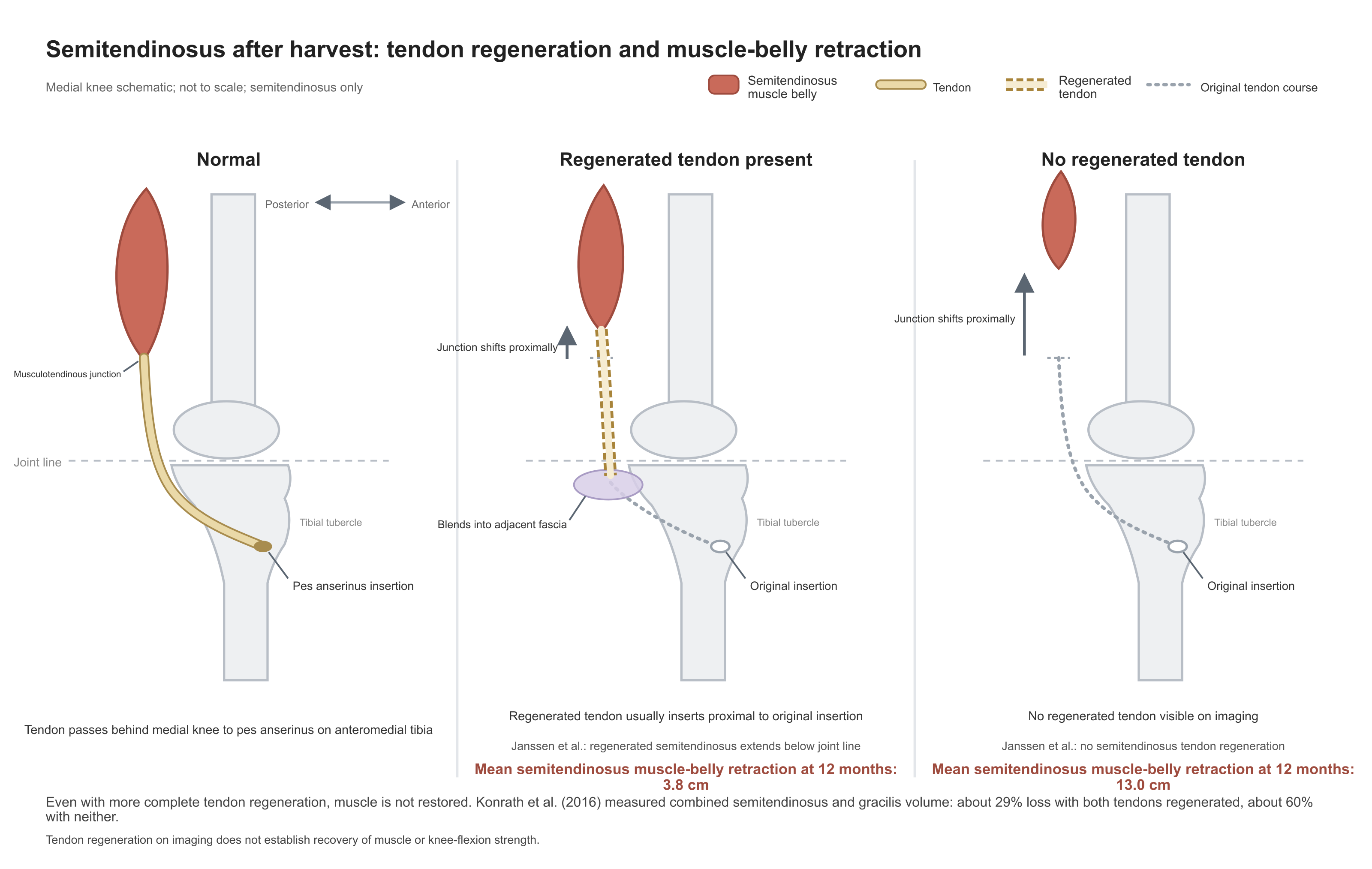
These are observational groupings; regeneration was not randomly assigned. Janssen et al. have preoperative imaging and can confirm that reduction occurred postoperatively, but cannot exclude the possibility that the severity of harvest injury, the extent of harvest, or surgical technique affected both regeneration and muscle retraction at the same time: people with less harvest injury and less muscle retraction may also find it easier for new tendon to grow back along the original path. What the available material can confirm is that more complete regenerators have less retraction and retain more muscle; it cannot confirm that regeneration preserved the muscle. Even more complete regenerators do not recover original muscle: in Konrath et al., those with both tendons regenerated still had about a 29% volume deficit of semitendinosus plus gracilis. Snow et al. at 9–11 years still found clear atrophy and fatty infiltration in the whole group (mean Goutallier grade 2.1 for semitendinosus, 1.8 for gracilis), without reporting by regeneration status.
Semimembranosus and Biceps Femoris
Whether the semimembranosus and biceps femoris enlarge to compensate after semitendinosus reduction is inconsistent across studies. Some see small increases of about 4%–10% in semimembranosus or long-head biceps volume; after meta-analytic pooling there is no difference:
| Study | Semimembranosus | Long head of biceps femoris | Short head of biceps femoris |
|---|---|---|---|
| Snow et al. (2012), 10 cases | No significant difference; no figures given | Volume 108.3% of contralateral (P=0.036; not significant after the authors' multiplicity correction); smaller in 2 | No significant difference; no figures given |
| Konrath et al. (2016), 20 cases | Volume +4% | Volume +7.7%, cross-sectional area +10.6% | — |
| Messer et al. (2020), 14 cases | Volume +9.7 cm³ (P<0.001) | No significant difference | Volume +5.9 cm³ |
| Hunter et al. (2026), 10 cases | 224.0 vs 194.9 cm³ (P=.106); 42.5% vs 36.6% of the posterior compartment (P=.006) | Not significant | Not significant |
| Sherman et al. (2021a), meta-analysis | No significant difference in volume or cross-sectional area | No significant difference | No significant difference |
Hunter et al. (2026) studied 10 subjects each with hamstring, patellar tendon, and allograft grafts, with a single postoperative MRI; mean time after surgery was about 7.5 years in the hamstring group (about 6.3 years in the other two, original Table 1). Mean absolute semimembranosus volume on the operated side in the hamstring group was about 15% larger than contralateral, P=.106, not significant; after conversion to percentage of posterior-compartment volume the difference was significant (original Table 3). On that basis the authors write of "compensatory hypertrophy of the semimembranosus" in the Abstract conclusion and Discussion. Significance of a proportion metric does not equal enlargement of absolute volume; with 10 per group and no preoperative imaging, even a side-to-side difference is hard to attribute to harvest. On the available data, compensatory hypertrophy can only be regarded as unconfirmed.
The P value for absolute volume has a further problem. On p. 6 of the Results the text gives 223.98±21.3 and 194.88±19.5 cm³ without stating whether 21.3 and 19.5 are SDs or SEs; Table 3 lists only means and P values, and the "mean±SD" footnote to Table 1 belongs to the demographic table. Recalculated from the original numbers, if those figures are SDs, a paired t test yields a maximum P of about 0.0506 no matter how low the between-side correlation, and an independent-samples t about 0.005—neither gives .106. If they are SEs, a between-side correlation of about 0.69 yields a paired P of .106. The Methods state only that the proportion metric used a paired t test and do not say what test was used for absolute volume. Until the authors clarify these two points, whether the absolute volumes differ cannot be judged from the available numbers. The hamstring group also has a numeric mismatch: the sum of the four posterior muscles on the operated side is 525.1 cm³, whereas Table 3 lists the posterior compartment as 537.6 cm³; the contralateral four-muscle sum matches the listed value (calculated from the original table), and the operated-side discrepancy is unexplained.
Table 2 and the text disagree for the allograft group and can be checked directly in the original: semimembranosus is 208.70/202.31 cm³ in Table 2 and 19.80/20.54 cm³ on p. 5; semitendinosus is 80.05/82.40 cm³ in Table 2 and 27.09/26.89 cm³ in the text. Detailed allograft figures cannot be used.
Two studies have preoperative–postoperative pairing, and the authors' wording differs. Williams et al. (2004), at return to sport in 8 subjects, report that semimembranosus volume and cross-sectional area exceeded preoperative values and that the long head of biceps femoris increased most, to 104% of preoperative volume and 105% of cross-sectional area, and state that both compensated (original Discussion); the Results report a significant muscle-group × side × time interaction but attribute that interaction entirely to reduction of the semitendinosus and gracilis and do not report significant increases in semimembranosus or biceps femoris. Janssen et al. (2013) found no significant compensatory hypertrophy of semimembranosus or biceps femoris at 12 months postoperatively (original p. 901) and give no cross-sectional area figures for those two muscles. Neither provides evidence of significant enlargement of the remaining hamstrings in the first half year to one year after surgery; the figures of Williams et al. show that they roughly retained preoperative size.
The author's judgment is that cross-sectional studies without preoperative imaging may be seeing increases that already existed before surgery; the amount of any increase falls far short of offsetting loss of the semitendinosus and gracilis—total hamstring volume in Messer et al. remained 9% below contralateral, and total hamstring-plus-gracilis volume in Williams et al. remained 10%–15% below preoperative. Confirming whether the remaining muscles enlarge requires preoperative–postoperative paired MRI that reports absolute volumes of each muscle.
Semitendinosus Participation During Movement
How much the atrophied semitendinosus still contributes during movement can only be estimated indirectly. Magnetic resonance T2 after exercise rises in rough proportion to how much a muscle has participated. Three T2 studies look contradictory, but their exercise tasks differ:
| Study | Population | Exercise task | Semitendinosus T2 rise |
|---|---|---|---|
| Takeda et al. (2006) | 11 cases, mean 12.7 months postoperatively | Alternating knee extension and flexion on an isokinetic dynamometer, 120°/s, 10 sets; body position not stated | Operated 10.9±2.7 ms, contralateral 10.6±3.2 ms, no difference; largest rise among the hamstrings on both sides |
| Messer et al. (2020) | 14 cases, median 49 months | Nordic eccentric hamstring exercise, 5 sets × 10 | Operated side 9.9 percentage points less than contralateral, about one third less, but still elevated; no side-to-side difference for semimembranosus or biceps femoris |
| Tampere et al. (2021) | 23 male football players at return to sport (about 10 months postoperatively) | Inclined-board prone leg curl to exhaustion | Operated 19.97%, healthy controls 25.32%; long head of biceps femoris operated 13.92%, healthy controls 8.48% |
The corresponding author of Messer et al. is a co-inventor of the patented Nordic device used and a minority shareholder in the manufacturing company. The T2 values in the table come from MRI before and after exercise and do not pass through that device; available materials do not show that this interest affected the T2 results.
Tampere et al. had subjects lie prone on a training table inclined 60°; the authors state that this posture flexed the hips about 60°, with both knees alternating between 90° of flexion and full extension, each foot loaded with 5 kg (original pp. 1473–1474). Of 30 patients originally recruited, 18 also had a lateral extra-articular tenodesis and 4 had additional gracilis harvest; how many of the final 23 fell into each category is not given, so the operated-side T2 change cannot be attributed entirely to semitendinosus harvest. For the 7 not analyzed, the Results text refers only vaguely to imaging problems and exclusions, whereas the flow diagram (original Fig. 4, p. 1475) lists rehabilitation nonadherence in 3, scan-image problems in 2, and hamstring injury before testing in 2. Operated-side semitendinosus T2 rise is 19.97% in the between-group analysis versus healthy controls (p. 1475) and 17.31% in the within-subject comparison with the contralateral side (p. 1477); how the two figures correspond is not explained. One author holds a patent licensed to Smith & Nephew; the patent content is not described, and any relation to this study is unclear. Lesser elevation of the semitendinosus is consistent with Messer et al.; greater elevation of the long head of biceps femoris appears only in this study.
In all three studies operated-side semitendinosus T2 rose after exercise, so the muscle still works after harvest. The Nordic exercise keeps the hip neutral while the knee moves from flexion to extension, and Tampere et al.'s leg curl flexes the hip about 60°; both include load with the knee near extension and the hamstrings at longer lengths, and operated-side semitendinosus elevation is smaller. Takeda et al.'s alternating isokinetic flexion–extension does not state body position and shows no side-to-side difference. Neither of the two exercises that show a difference used hip extension with deep knee flexion and short hamstring lengths; contraction mode, population, and T2 calculation also differ. Neither study measured deep-flexion strength; under which muscle lengths semitendinosus participation falls most, and whether that relates to the deep-flexion strength deficit, requires studies that measure both T2 and deep-flexion strength.
The title of Einarsson et al. (2021) states that medial hamstring activity during running is lower after ACL reconstruction. The study compared medial (semitendinosus) and lateral hamstring EMG on a treadmill in 21 male ACL-reconstructed athletes (hamstring graft 12, patellar tendon 9, 5–9 months postoperatively) and 19 healthy runners. EMG was normalized to each person's own peak during running; all tests listed in Table 1 are within-group comparisons of swing versus stance (original pp. 2–3). The Methods mention ANOVA but give no between-group results that would support "lower in the hamstring-graft group than in the patellar-tendon group or in healthy people"; controls were also about 9 years older on average than patients, so even a between-group comparison would be hard to attribute to harvest. This normalization itself cannot compare absolute activation across people.
The original has further inconsistencies. The Results text says medial hamstring integrated EMG was lower in swing, whereas Table 1 shows it lower in stance; the contralateral peak comparison in the hamstring-graft group has P=0.06, yet the Abstract says all conditions were significant. In Table 1 the operated-side medial integrated EMG row for the patellar-tendon group is 45.27±16.54 and 10.03±4.65, and the contralateral row for the hamstring-graft group is 45.27±16.54 and 10.03±4.64—identical except for the last digit; lateral peaks on both sides in the hamstring-graft group are also identical, unexplained. What Table 1 can confirm is only the within-group phase distribution: medial hamstring was relatively lower in stance in every group. That is a within-person comparison across phases and says nothing about absolute semitendinosus activation after harvest.
Modeling Studies
Konrath et al. (2017) and du Moulin et al. (2026) use musculoskeletal models to conclude that other muscles share load. The former fed literature atrophy parameters for semitendinosus and gracilis into a model; their contribution to medial tibiofemoral compartment load fell 17%–26%, with a corresponding rise in the semimembranosus contribution. The latter simulated sidestep cutting with MRI morphology from 18 subjects in the Konrath cohort and likewise saw load shift toward the semimembranosus and long head of biceps femoris.
Both models require reproduction of the same set of measured joint moments. When one muscle's capacity shrinks and total moment is held constant, the calculation redistributes load to other muscles; how much each receives depends on the model's constraints and optimization rules. So-called compensation is the model redistributing load to other muscles under given constraints; neither paper measured increased force output from other muscles.
The two papers word their conclusions differently. The Abstract conclusion of Konrath et al. (2017) states that the semimembranosus "can compensate" (original p. 2); the Limitations acknowledge that atrophy parameters were literature averages rather than individual measured morphology, and the body conclusion softens to "appears possible" (p. 16). The Abstract and body conclusion of du Moulin et al. (2026) (p. 8) both state that persistent impairment of semitendinosus function "leads to" compensatory redistribution of hamstring load, reading between-muscle load differences in the model directly as compensation; the Limitations (p. 7) acknowledge that static optimization assumes an ideal minimum-activation pattern that does not reflect individual neural control and may not suit high-intensity actions such as cutting, and the study did not directly measure each muscle's force output.
4. Knee Flexion Strength
How much knee flexion strength falls after harvest depends on the conditions of measurement. In the same patients at the same testing session, strength at conventional or shallow angles can approach the contralateral side while torque with the hip extended and the knee in deep flexion remains clearly deficient; representative deep-flexion LSI values across studies mostly lie between about 50% and 80% of the contralateral side.
Testing Position and Muscle Length
The semitendinosus, semimembranosus, and long head of biceps femoris all cross both the hip and the knee. Hip flexion and knee extension lengthen them; hip extension and knee flexion shorten them. The same label "knee flexion strength" therefore implies different muscle lengths depending on hip and knee angles. Common tests fall into three conditions:
- Seated isokinetic testing: hip flexed about 90°, hamstrings at a longer length.
- Prone or supine deep-flexion testing: hip extended and knee flexion deepened, the shortest position of the hamstrings. In this chapter "deep flexion" means a larger knee-flexion angle, usually 90° or more. Seated tests also include deep-flexion angles (for example the angle curves of Hart et al. below), but the hip remains flexed and the hamstrings are not as short.
- Nordic testing: hip held neutral, knee moving from flexion to extension, hamstrings gradually lengthened under eccentric contraction, both legs producing force together. Hip position and contraction mode both differ from seated testing; the gap between the two results cannot be attributed to either factor alone.
Most studies use a seated isokinetic dynamometer, flexing the knee at 60°/s or faster and recording the maximum torque over the movement (peak torque). Peak torque usually occurs at about 20°–40° of knee flexion: Ogborn et al. (2023) measured 36° on the operated side and 39° on the contralateral side when seated, and Vairo et al. (2014) about 30° when prone. By the architectural inference of Section 1, the semitendinosus share is modest in this range. Strength measured under the three conditions is not interchangeable. Representative post-harvest results under each condition are as follows:
| Testing condition | Hamstring length and contraction mode | Representative results after harvest |
|---|---|---|
| Seated isokinetic peak torque | Longer, concentric | At 60°/s flexion, group-mean LSI 89.0% at 12 months and 91.7% at 24 months postoperatively (Högberg et al., 2024a); compared concurrently with other grafts, the hamstring group is usually 7–15 percentage points lower |
| Deep flexion (mostly prone, hip extended, knee flexed 90° or more) | Shortest | Representative LSI values across studies mostly 49%–80% (see deep-flexion strength table below) |
| Nordic eccentric | Gradually lengthened, eccentric | Mean LSI 79%–90% (Ogborn et al., 2023; Högberg et al., 2022, 2024b); at 12 months postoperatively, LSI ≥90% in 73% on seated testing and 41% on Nordic testing (Högberg et al., 2022) |
| Internal tibial rotation at 90° of knee flexion | — | About 12%–15% lower after semitendinosus plus gracilis harvest; the author's estimate from the measured values of Armour et al. (2004) and Konrath et al. (2016) |
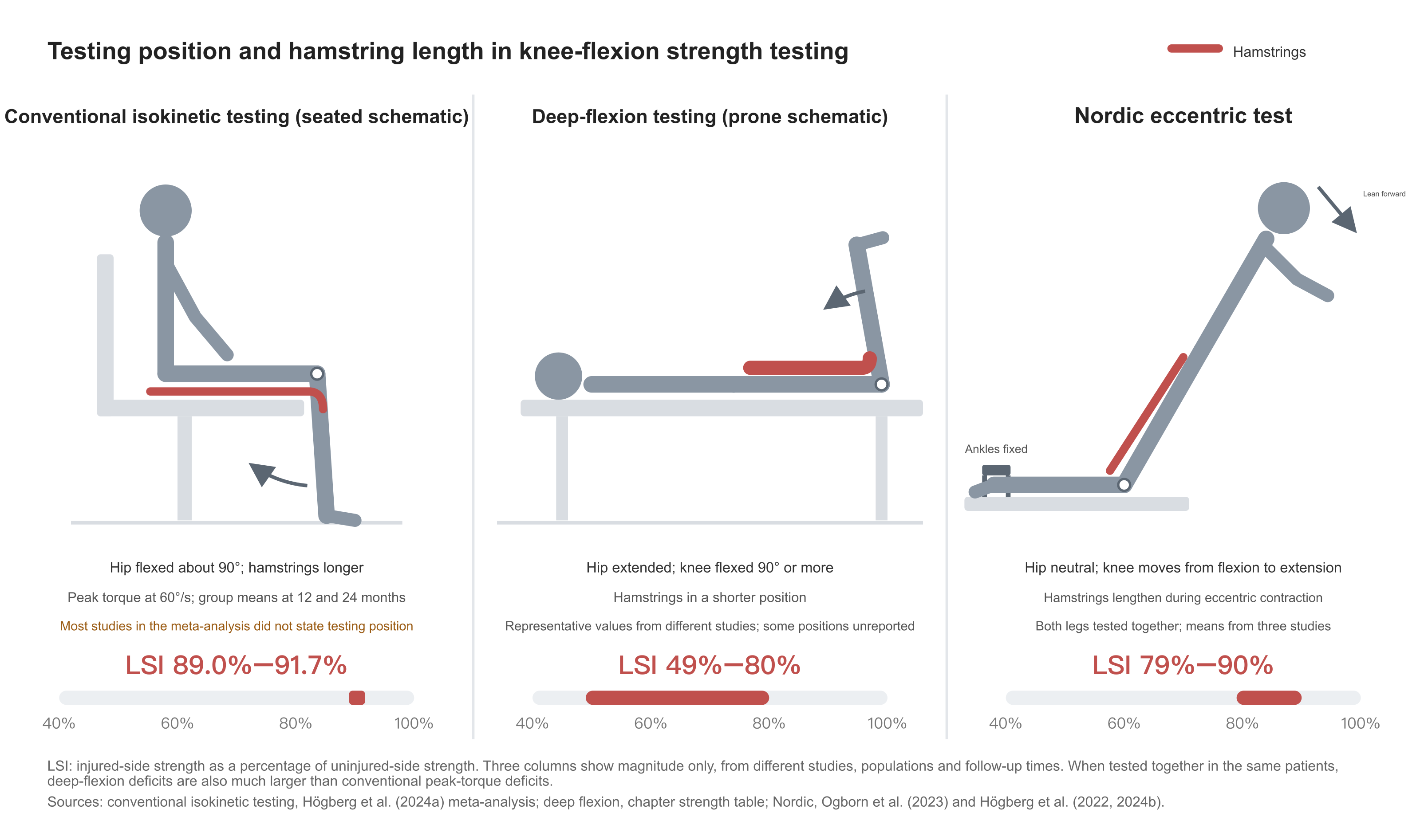
Among comparisons that change only one testing condition in the same people, Ogborn et al. (2023) come closest to a single variable. Thirty-one subjects with semitendinosus plus gracilis harvest about 14 years postoperatively all performed 90°/s concentric isokinetic testing: peak-torque LSI was 95.8% seated (hip flexed 70°–85°) and 93.8% supine (hip 0°); at 60° of knee flexion, seated 93.6% and supine 82.7%; at 75° supine, 71.1%. Nordic eccentric testing was 83.9%; the difference from seated peak torque was not significant (original Results and Fig. 1).
Between seated and supine only the hip angle changed. Each leg was always tested seated first, so order effects cannot be excluded; the authors did not directly test the seated–supine difference at 60°, and 93.6% and 82.7% are simply two juxtaposed figures. Hip extension used the supine position; most deep-flexion studies use prone. Nordic testing, beyond the differences already noted, also changed the testing device. Only 31 of 203 subjects in the original database (15%) completed all tests. The size of the differences can therefore be used only roughly; the direction that deficits are larger with the hip extended at the same knee angle is consistent with Koutras et al. below and with the deep-flexion data.
Koutras et al. (2016), in 42 cases on the same machine at the same speeds, compared two positions: the flexion deficit measured prone (hip 0°) averaged 6.5 percentage points larger than seated (hip 90°) at 60°/s and 9.1 at 180°/s, and remained 4 and 9 percentage points larger at 9 months postoperatively. Values at each time point appear only in bar graphs. The Abstract states that "most athletes could not be cleared at 9 months"; the text does not report individual pass rates, so that sentence lacks a data basis; the mean between-position difference is a measured result.
Two studies from the same Swedish rehabilitation registry compared seated isokinetic with Nordic testing. Högberg et al. (2022), at each time point in the first postoperative year, found mean seated flexion LSI 88%–95% and mean Nordic eccentric LSI 79%–87%, a gap of about 8–13 percentage points. Högberg et al. (2024b), in two different patient groups at 2 and 5 years postoperatively, found mean seated LSI about 99%–101% and Nordic 86% and 90%. The two papers may share patients; the originals do not give the intersection and cannot be treated as two independent samples.
Group Means on Conventional Testing
The most complete group means come from the meta-analysis of Högberg et al. (2024a). Pooling 64 studies, 60°/s flexion LSI was 80.8% preoperatively, 89.0% (95% CI 87.3–90.7) at 12 months postoperatively, and 91.7% (90.8–92.6) at 24 months, with low or very low certainty of evidence. The 12- and 24-month figures come from almost nonoverlapping sets of studies (24 months mainly several Japanese studies); the two time points are a spliced cross section and cannot be read as the same people recovering another about 2.7 percentage points in the second year; the authors also regard the clinical meaning of the 89% versus 92% difference as doubtful. The legend to their Figure 4 misprints the 6-month values as the 12-month values. Most included studies in this meta-analysis do not state testing position (original pp. 11, 13), which the authors themselves list among uncertainties in interpreting the results. A mean 60°/s flexion LSI of about 90% at one year postoperatively is the order of magnitude from studies that mainly used conventional testing with position often unstated; it does not represent deep-flexion or Nordic deficits.
LSI uses the contralateral side as reference, and that side may also weaken during injury, surgery, and rehabilitation. Hiemstra et al. (2007), in 12 cases (9 isolated semitendinosus) about 3.4 years postoperatively, found contralateral flexion strength below healthy controls: the Abstract gives 13.5% lower; averaging across speeds from their Table 3 gives 11.6%. All 30 controls were men, whereas the patient group included 5 women, so part of the gap may be sex. In the randomized trial of Sinding et al. (2020), the contralateral side in both patient groups matched age- and sex-matched healthy controls. Whether the contralateral side weakens is inconsistent across studies. Looking only at LSI may overestimate recovery; reporting body-weight-normalized absolute torque (Nm/kg) as well is more robust.
The large meta-analysis of Girdwood et al. (2025) (232 studies) states in the Abstract that flexion was only 5%–7% down at 1 year. It pools all graft types; hamstring grafts were only 33%, and 46% of studies did not state the graft. Concentric testing at 1 year was about 94%, isometric only 84.5%; the Abstract folds isometric into the "5%–7%" as well, which is wrong. The Discussion states that "by 5 years strength had returned to the level of the contralateral side or healthy people," yet Table 1 gives concentric strength at 5 years as 92.9% (90.8–95.1), a confidence interval that excludes 100%; that sentence likewise does not hold. The only hamstring-specific result in Girdwood et al. comes from a sensitivity analysis: flexion strength in the hamstring group was 0.92 times that of other graft groups (slow concentric) and 0.84 times (isometric). These ratios can show that the hamstring group is weaker than other graft groups; they should not be used to estimate how far harvest patients themselves recover. The Abstract's "5%–7%" mixes all graft types and should not be cited as the post-harvest deficit for hamstring tendon.
Figures from the Swedish registry in Högberg et al. (2026) are high. In 406 hamstring-graft patients, flexion LSI was 89.6% at 4 months, 95.3% at 8 months, and 97.5% at 12 months. The study included only patients who completed all three tests at 4, 8, and 12 months; of 687 who met inclusion criteria, a further 167 (24%) were excluded for two or more cumulative ACL injuries (ipsilateral or contralateral); contralateral strength in the hamstring group was itself lower than in the patellar-tendon group (1.40 vs 1.53 Nm/kg at 4 months), so the LSI denominator is small; confidence intervals in several tables are misprinted. These means reflect patients who completed all three tests and had only one cumulative ACL injury; the printed confidence intervals should not be cited.
Single-center small-sample results are much lower. Burks et al. (2005), in 9 cases of semitendinosus plus gracilis harvest, found 60°/s peak flexion force 21% (7%–39%) below contralateral at 12 months and 13% below at 180°/s, the latter not significant; 2 of 9 had poor rehabilitation adherence, so the mean is heavily influenced by individuals.
Comparison with Other Grafts and with the Unharvested Leg
In concurrent comparisons with other grafts, seated flexion LSI in the hamstring group is usually 7–15 percentage points lower:
| Study | Design | Hamstring group | Comparator graft | Flexion LSI (hamstring vs comparator) |
|---|---|---|---|---|
| Cristiani et al. (2021) | Randomized trial, 160 cases | Semitendinosus plus gracilis | Patellar tendon | 90°/s: 79.5% at 4 months, 87.4% at 12 months, 89.1% at 24 months; patellar-tendon group 97.1% at 24 months (original Fig. 3a) |
| Sinding et al. (2020) | Randomized trial, 100 cases | Semitendinosus plus gracilis | Bone–quadriceps tendon | At 1 year, 60°/s: 81.3% vs 95.9%; healthy controls 96.1% (original Table 3) |
| Ebert et al. (2024) | 116 cases | >90% isolated semitendinosus | Soft-tissue quadriceps tendon | 87.7% vs 95.5% at 6 months; 94.0% vs 101.1% at 24 months |
| Popovic et al. (2024) | Randomized 114 cases; strength measured in 61 | Hamstring 33 | Patellar tendon 28 | At 18 years, 60°/s: 90.6% vs 100.4%; total work 86.4% vs 101.3% |
Preoperative flexion LSI in both groups of Cristiani et al. was only about 84.5%; asymmetry already existed after injury, so the postoperative deficit cannot be attributed entirely to harvest. The trial enrolled in 2002–2005 and was published in 2020; at 24 months only 106/160 (66%) had data. Sample size was calculated on a difference in kilopond-metres, not the reported LSI, so the trial's power for between-group LSI differences cannot be judged.
In Ebert et al., neither assessors nor patients were blinded, and rehabilitation was completed by patients individually in the community; the hamstring group mainly had isolated semitendinosus harvest, unlike the other double-tendon harvest studies in the table. Assessors in Popovic et al. were unblinded. The text's "flexion strength 10.7% lower" is a between-group ratio with the hamstring group as denominator and is not the same as a side-to-side deficit; the LSI difference between groups is 9.8 percentage points. Patient-reported outcomes, KT-1000 laxity, and osteoarthritis did not differ detectably between groups.
The network meta-analysis of Liu et al. (2026) pools randomized trials and observational studies; flexion LSI was about 9 percentage points higher with quadriceps tendon than with semitendinosus plus gracilis (9.20 at 60°/s, 9.28 at 180°/s); the 4.12-point difference between patellar tendon and semitendinosus plus gracilis was not significant; certainty of evidence was very low to low. It reports no significant differences among grafts at 18 months or later, yet at 18 years in Popovic et al. the two groups still differed by 9.8 LSI points.
All of the above comparisons mix the effects of harvest and of reconstruction. Designs that harvest from the uninjured side and reconstruct the injured side can isolate harvest itself. Yasuda et al. (1995) alternately allocated 70 cases; the leg that was harvested but not reconstructed had 60°/s peak flexion torque at 12 months equal to 99% of preoperative, while the contemporaneous unoperated leg rose to 122% (original Table 8). The authors judged the effect "minimal" against the preoperative baseline; against the contemporaneous control, the harvested leg was about 23 percentage points lower (calculated from the original table). In the randomized trial of von Essen et al. (original unread; cited via Vivekanantha et al., 2023), contralateral-harvest legs had lower 60°/s flexion strength at 6 months than unharvested legs (91.4 vs 111.5; units not stated in the review's Table 3; P=0.001), and the difference was no longer significant at 24 months. Both studies measured only conventional-speed peak torque; there are no deep-flexion data.
Individual Pass Rates
A group mean LSI of about 90% still leaves large differences among individuals. Studies that directly report individual pass counts are few and all come from the Swedish rehabilitation registry. Högberg et al. (2022), 41 subjects at each time point: at 12 months, seated isokinetic flexion LSI ≥90% in 30 (73%) and Nordic in 17 (41%). Högberg et al. (2024b), 48 subjects each at 2 and 5 years postoperatively: seated pass rates both 42 (88%), Nordic 19 and 23 (40% and 48%). By these figures, about one quarter fail to reach 90% on seated testing at one year postoperatively, and more than half fail on Nordic testing.
Both studies included only people with high preinjury sport level (Tegner ≥6) who had performed Nordic exercise during rehabilitation; how the 41 at each time point were selected is not stated. Sundberg et al. (2025) from the same registry likewise included only preinjury Tegner ≥6; of 526 cases at return to sport (mean about 9.6 months postoperatively), 376 (71.5%) had seated 90°/s flexion LSI ≥90% (original Table 1; see Section 9). Direct data on individual pass rates outside the registry are absent from the available material.
Högberg et al. (2022) state in the Methods that pass rates were compared with Fisher's exact test, yet recalculation with that test (from the original table counts) does not reproduce the printed P values at any of the four time points (original Table 3). The two tests were in the same people and should use a paired design; exact McNemar can reproduce the 4- and 12-month P values, but not those at 10 weeks and 8 months. Whichever test is used, seated pass rates exceed Nordic at all four time points and the differences are significant, so the direction of the conclusion is unchanged; the printed P values at 10 weeks and 8 months cannot be reproduced and should not be cited.
Deep-Flexion Strength
In the same patients at the same testing session, conventional peak torque approaches the contralateral side while deep-flexion torque remains clearly deficient. The deeper the angle, the larger the deficit—a finding repeated across countries, testing devices, and both isolated semitendinosus and double-tendon harvest, and the most consistent direction in the post-harvest strength literature.
| Study | Harvest | Follow-up | Conventional or shallow angle | Deep flexion |
|---|---|---|---|---|
| Nakamura et al. (2002) | Semitendinosus 49, plus gracilis 25 | 2 years | Peak-torque LSI 91%–94% | 72%–80% at 90° on the same curve (position not stated) |
| Tadokoro et al. (2004) | Semitendinosus plus gracilis 28 | Mean 5.6 years | Seated 90° isometric 86.2% | Prone 90° 54.6%, prone 110° 49.1% |
| Nishino et al. (2006) | Isolated semitendinosus 23 | Mean 23 months | Prone 45° 94.1% | Prone 90° 74.0% |
| Makihara et al. (2006) | Semitendinosus 12, plus gracilis 4 | Mean 26 months | Isokinetic peak torque about 96%; prone 45° isometric no difference | Prone 90° isometric: operated 55±19, contralateral 88±28 (% body weight) |
| Åhlén et al. (2012) | Semitendinosus plus gracilis 19 | Median 8.5 years | Not reported | Torque at prone 90° of flexion 69% (60°/s), 78% (180°/s) |
| Nomura et al. (2015) | Isolated semitendinosus 24 | 12–72 months | Prone 30° about 91% | Prone 90° about 68%, 105° about 69% |
| Kositsky et al. (2024) | Isolated semitendinosus 10 | About 14 months | Operated below contralateral: about 6% at 15°, about 10% at 45° (neither significant) | Prone, operated below contralateral: about 22% at 60°, about 32% at 90° |
| Vairo et al. (2014) | Semitendinosus plus gracilis, 15 women | About 26 months | Prone full-range peak torque 0.959 vs 1.012 Nm/kg | At prone 105° 0.221 vs 0.400 Nm/kg (conventional LSI about 55%) |
Percentages not otherwise noted are LSI. The 90° values of Nakamura et al. come from isokinetic torque curves; the original does not state position. Deep-flexion testing in the remaining studies was prone (hip extended).
The "53.83% deficit" in Vairo et al. uses a symmetry percentage difference with the two-side mean as denominator; by the conventional algorithm the operated side is about 55% of contralateral. Retest reliability of torque at 105° was low (ICC 0.695, lower confidence limit 0.093); group means can be used, but individual values fluctuate widely and should not be used to judge a single patient. The "±" in Table 2 of Kositsky et al. is the half-width of the 95% confidence interval; reading it as an SD underestimates between-person dispersion.
Makihara et al. always tested the contralateral side first; when the operated side was tested later the subject already knew the test, and any learning effect would bias operated values upward and shrink the deficit. Of 16 subjects, 13 were women. Studies in the table are all cross-sectional and small-sample; most lack preoperative data.
Hart et al. (2022) reached the same result with full torque-curve analysis. Three hundred fifty-seven male field-sport athletes underwent seated isokinetic testing 8–10 months postoperatively; 60 had hamstring grafts (single or double tendon not reported). Peak flexion torque asymmetry was 17% in the hamstring group and 10% in the patellar-tendon group; looking at the largest asymmetry over 15°–85°, the hamstring group reached 36% at about 56° of flexion and the patellar-tendon group 29% at about 47° (original Table 2). Peak torque underestimates flexion asymmetry, and the largest deficit in the hamstring group occurs at a deeper angle. Grafts were not randomly assigned; the hamstring group was lighter.
The curve analysis of Hart et al. also gives peak effect sizes: extension asymmetry 5.53 at 75° of flexion, flexion asymmetry 2.68 at 85°. The original does not report means and SDs at these two angles, so the effect sizes cannot be checked; the author does not use them to judge the size of between-group differences. The legends to Figs. 1 and 2 label the between-group difference in asymmetry as "Nm/kg"; the percentage values in Table 2 are unaffected by that label.
The angle of peak torque and the standing active flexion angle also change with how much tendon is harvested; those data (Adachi et al., 2003) are discussed with the single- versus double-tendon comparison in Section 5.
Internal Tibial Rotation and Hip Extension
The semitendinosus and gracilis are both internal rotators of the tibia (Section 1). Three studies found internal-rotation strength at 90° of knee flexion below the contralateral side after semitendinosus plus gracilis harvest, with external rotation unaffected. From the percentages of Armour et al. and Konrath et al., the author estimates the fall in internal-rotation strength at 12%–15%; Viola et al. point in the same direction.
Armour et al. (2004), in 30 cases at least 2 years postoperatively, found internal rotation 12%–15% lower (original Table 2); the authors treated the two sides of the same person as two independent samples, a conservative test. Konrath et al. (2016), supine at 90° of flexion and 60°/s, found internal rotation about 14% lower. Viola et al. (2000), in 23 cases at a mean of about 4 years postoperatively, found contralateral-minus-operated peak internal-rotation torque of 2.6–3.2 ft-lb, P≤0.0001 at each speed; the overall difference at 180°/s weighted from the sex-stratified tables should be 3.9 (recalculated from the original numbers), which does not match the 3.2 in the text—the text figure is too small, but the conclusion of weakened internal rotation is unaffected.
Åhlén et al. (2012) point in the opposite direction: at 8.5 years, operated-side internal rotation at 60°/s was stronger (43.0 vs 36.7 Nm). That study always tested the contralateral side first and the operated side second; testing order may have biased operated values upward.
Hip-extension data are scarce. Geoghegan et al. (2007), in 10 cases of semitendinosus plus gracilis, found concentric peak hip-extension force on the operated side 0.89 times contralateral at 3 months postoperatively, versus 1.13 in a patellar-tendon group; at 12 months they report "no significant difference" without giving figures; the groups differed in sex and age, and grafts were chosen by sport. Hadi et al. (2019), in 20 cases of semitendinosus plus gracilis, found the two sides similar at 3 months. Both are small samples; one can say only that hip-extension strength may fall slightly early on, with a lack of data at 1 year.
Eccentric Strength and Rate of Force Development
Nordic eccentric results in athlete studies disagree in direction. Bourne et al. (2019), in 12 elite Australian-rules football women with a history of semitendinosus harvest at a median of 4.5 years postoperatively, found Nordic eccentric peak force about 54 N (about 20%) lower on the operated than the contralateral side. Messer et al. (2022), in male Australian-rules football, soccer, and rugby athletes, found that among 43 with a history of ACL reconstruction and no recent hamstring strain, Nordic eccentric force differed little from uninjured athletes (−15 N, P=0.646); graft type was unrecorded in most. The former compares sides; the latter compares with uninjured people; sex, sport, and graft recording also differ, so the divergent results cannot be attributed.
Rate-of-force-development data come from San Jose et al. (2023). In 89 hamstring-graft patients, from 3–6 to 7–12 months postoperatively, seated concentric flexion LSI rose from 86% to 92%, isometric maximum force at 45° of flexion from 76% to 84%, and isometric rate of torque development (reflecting explosive strength) changed from 86% to 83%, a nonsignificant change. The title states that "maximum strength has recovered while explosive strength has not," yet isometric maximum strength was likewise only 84%; individual variation in rate of torque development was extreme (SD 22–49), and only 42 subjects took the later test. These data can show that both isometric indices remain clearly below the contralateral side within the first postoperative year; whether explosive strength recovers more slowly than maximum strength, they cannot answer.
Sources of the Deficit
The available evidence points to several possible explanations for the deep-flexion deficit: semitendinosus shortening, a reduced moment arm after proximal migration of the insertion, and reduced force capacity of the shortened muscle at short muscle lengths. Support for these explanations is cross-sectional correlation, cadaveric passive measurement, and model calculation; the three classes of evidence cannot be strung into a confirmed causal chain. Changes in neural control and intramuscular architecture may also contribute; none of the studies cited here measured them directly, and the share of each factor has not been measured.
Konrath et al. (2016) found that volume, cross-sectional area, and length deficits of semitendinosus plus gracilis correlated with the flexion strength deficit (r=0.51, 0.57, 0.61); morphology explained only about a quarter to a third of the variance (by r²).
Moment-arm data come from cadavers. Snoeck et al. (2021), in 7 cadaveric lower limbs, first harvested the gracilis and then the semitendinosus; the peak of the semitendinosus flexion moment-arm curve fell to about 61% of intact (27/44 mm), with the largest drop at 80°–90° of flexion (38→11 mm); cutting the insertion without stripping left the moment arm unchanged (104%). These are passive, nearly unloaded measurements; a smaller moment arm does not mean that torque falls by the same proportion. The data are a reanalysis of that team's 2014 study. The Abstract states P≤0.005; Table 4 gives P=0.029 for the intact-versus-post-harvest comparison, still below 0.05, so the conclusion of reduced moment arm stands, though the Abstract overstates the level of significance.
du Moulin et al. (2024) used individualized models of 7 subjects whose semitendinosus had regenerated to the pes anserinus level, drawn from the Konrath cohort. The model calculated an "asymmetry index" of 19.32% for single-muscle semitendinosus flexion torque, 15.5% for internal rotation, and about 6% for whole-hamstring flexion. The denominator of this index is the sum of the two sides; 19.32% corresponds roughly to the operated side being 68% of contralateral. It computes "torque capacity" assuming full muscle activation, not measured strength, and cannot be written as "flexion strength falls 19% after harvest." The authors acknowledge that the model did not reproduce the measured deep-flexion deficit (Discussion, third paragraph); the Discussion also writes the index as 19.6%. Three authors disclose participation in an Australian Research Council Industrial Transformation Training Centre in collaboration with Arthrex; the study is model calculation and involves no specific device. The confidence intervals are also questionable: if the reported SD is the between-subject SD of 7 people, conventionally calculated confidence intervals should be much wider; the original does not state whether the interval used patients or angles as the unit, so it cannot be checked. That point affects only interpretation of precision, not the figures above.
In the model of Kositsky et al. (2024), shrinking only semitendinosus cross-sectional area had little effect at any angle; adding sarcomere-level shortening could explain the deficit at 60°; the deficit at 90° of flexion could not be explained by any morphologic change, and even a 20% enhancement of the other flexors could not make it up. This is a single generic model that depends on multiple assumed parameters.
Modifications That Preserve the Insertion
If the deficit relates to muscle retraction and proximal migration of the insertion, retaining part of the distal attachment may reduce the deep-flexion deficit. In the three studies that measured strength, the attachment-preserving group had better results on at least some indices, and the MRI of Low et al. also showed less muscle retraction; these are before–after period comparisons, retrospective comparisons, or small samples with incomplete between-group testing, and cannot yet show that preserving the attachment itself reduced the deficit.
Sasahara et al. (2014), comparing two successive periods, found that at 2 years the 20 cases with a preserved lateral 2 mm strip of semitendinosus shortened by 8 mm (0–40), versus 36 mm (10–70) in 16 fully harvested cases. Prone isometric LSI at 90° of flexion was about 87%–90% in the partial-harvest group and 55.3±13.9% in the full-harvest group (P<0.001); at 30° and 60° the groups did not differ.
Partial-harvest figures are 87.0±20.4% in the Abstract and 90.4±14.9% in the Discussion—inconsistent; both are far above the full-harvest group, so the direction of the between-group comparison is unaffected, but the exact partial-harvest value cannot be determined. Standing active flexion angle as a percentage of contralateral was 99.0% and 94.9% in the two groups; the original wrote "no significant difference (P=0.012)," and a 2015 correction changed that sentence to "significant difference" without changing the figures or correcting the 87.0% versus 90.4% inconsistency. The control was a nonrandomized successive period; 3 patients whose graft diameter after partial harvest was inadequate and who were converted to full harvest were excluded and not assigned to the full-harvest group, so the partial-harvest group may be biased toward people with better tendons; 2-year follow-up was 66%. The partial-harvest group had less shortening and better deep-flexion strength, but the between-group difference mixes selection bias and does not represent the effect size of the technique itself.
Ferretti et al. (2008), allocating by lot, found that 17 men with a preserved 4 cm distal semitendinosus stump had internal-rotation torque LSI at 90° of flexion of 97.4% at 25 months, versus 84.6% in 14 conventional cases. The groups were compared only within groups, without between-group tests or reported SDs, so whether the 97.4% versus 84.6% difference exceeds random fluctuation cannot be judged; 2 stump-group patients with tendons that were too short were also excluded after randomization. Low et al. (2024), on 6-month MRI in 30 cases, found proximal myotendinous migration of 3.4 cm in the group with a preserved 2 cm stump and 5.7 cm in the conventional group; strength was not measured.
Catma et al. (2024), in a retrospective comparison of 34 cases, found body-weight-normalized peak flexion torque at 180°/s of 0.68 vs 0.52 at 2 years in the group that preserved sartorial fascia attachment (P=.002), with no difference at 60°/s; the original labels the unit "%," yet the magnitude looks more like Nm/kg. Table 3's side-to-side "deficit" has no calculation formula; subtracting group means in Table 2 does not yield Table 3's figures; if it is the mean of each person's relative deficit, the two need not match. What can be confirmed is that the definition and unit of the deficit are unclear and cannot be recalculated from the summary data, so Table 3 cannot be used to compare side-to-side deficits between groups. The study was not randomized and did not measure deep flexion.
5. Semitendinosus Alone Versus Semitendinosus Plus Gracilis
Whether to add gracilis harvest when the semitendinosus is adequate requires weighing two things: how much more flexion strength is lost by taking a second tendon, and whether using only the semitendinosus yields a graft that is too thin or too short. On seated conventional peak torque the two approaches differ little; differences concentrate in deep flexion, internal tibial rotation, and active flexion angle. Studies that measured those outcomes have small samples, and most were not randomized.
Many comparative studies decide whether to add gracilis by semitendinosus thickness. Segawa et al., Nakamura et al., Adachi et al., Sengoku et al., and Cristiani et al. (2024) all added gracilis only when the semitendinosus was too thin or too short; patients in the double-tendon groups already had thinner tendons, and several studies also had clearly more women. Between-group differences may come from the gracilis, from body size, sex, or the graft itself. Grouping methods in the remaining studies vary and are noted with each study.
Whether the gracilis is truly intact after isolated semitendinosus harvest is poorly documented. Testory et al. (2022), in 50 adolescents with isolated semitendinosus harvest, saw the gracilis on MRI at the medial femoral epicondyle level in every case at 1 year postoperatively, with cross-sectional area rising from 7.1 mm² to 8.6 mm². That level is above the joint line and cannot show whether the distal gracilis was damaged; there was no control group, and the area increase may reflect growth. What can be confirmed is only that the main body of the gracilis remains after semitendinosus harvest; neither "isolated semitendinosus harvest spares the gracilis" nor "compensatory gracilis thickening" follows.
Conventional Seated Peak Torque
In several meta-analyses, seated peak-torque differences between single and double tendon are small and near zero at 24 months or later; early pooled results that reached significance sometimes depend on how the data were handled.
| Review | Included | Seated peak-torque results | Main problems |
|---|---|---|---|
| Sharma et al. (2016) | 12 studies; 4 labeled randomized trials | Single-tendon LSI minus double-tendon LSI: 3.85 percentage points at 60°/s (95% CI 0.77 to 6.92, 8 studies); 3.20 at 180–240°/s (95% CI −0.33 to 6.72, Abstract P=.08), yet the body writes 2.39% (P=.18)—neither significant | Abstract and body disagree at 180–240°/s; rotation-allocated studies labeled level-1 randomized trials; no certainty-of-evidence rating |
| Matteucci et al. (2024) | 15 studies (1227 subjects) | At 6 months, "all speeds pooled" standardized mean difference −0.25 (95% CI −0.40 to −0.10, P=.001) favoring isolated semitendinosus; at 24 months or later at 60°/s −0.10 (−0.26 to 0.07, 8 studies, I²=0%) | Data at different speeds from the same patients entered repeatedly; multiple inconsistent headcounts; Sengoku et al. used preoperative contralateral as the LSI denominator |
| Ardern and Webster (2009) | 7 studies | Conventional isokinetic peak torque did not differ between single and double tendon; deep-flexion and prone differences came from Tashiro et al. alone | Body claims 519 subjects total; table totals (556 or 481) match neither |
| Liu et al. (2026) | Network meta-analysis | States that "four-strand isolated semitendinosus may be superior to semitendinosus plus gracilis"; no comparison was significant (overall difference −6.63, 95% CI −15.55 to 1.88) | The conclusion rests only on ranking probabilities |
These reviews overlap heavily in included studies; agreement of conclusions largely reflects use of the same primaries.
Sharma pooled 4 "randomized trials" with 8 nonrandomized studies; one author discloses funding from Sanofi and Smith & Nephew. In Carter and Edinger (1999), labeled a level-1 randomized trial, all operations were performed by one surgeon; 120 cases were assigned by "every three cases in rotation" to patellar tendon, isolated semitendinosus, and semitendinosus plus gracilis (analyzed 106: 38, 33, 35)—rotation allocation—yet the Abstract writes "randomly selected" and the Methods "prospective randomized." It measured only 180°/s and 300°/s at 6 months postoperatively; single- versus double-tendon flexion LSI was 80.6% vs 81.7% and 79.1% vs 75.6%, with no difference. Sharma placed it in the 180–240°/s column and not in the 60°/s pool. Data extraction for this study was not wrong; the problem is labeling a rotation-allocated study as a level-1 randomized trial.
Matteucci et al. pooled only seated peak torque; the only result that reached significance in the full text is the 6-month "all speeds pooled" entry above. That pool enters different speeds from the same patients as independent rows; Figure 4 totals "353 vs 323 subjects," whereas the 4 studies actually comprise about 152 vs 148 (calculated from study headcounts). Figure 4 has two further headcount problems: the Sengoku et al. (2022) 180°/s single-tendon row lists 45, against 41 in the original; the Drocco et al. 240°/s double-tendon row lists 27, against 45 in the other rows of that study in the same figure and in review Table 1—internal inconsistency.
The heavily weighted Sengoku et al. (2022) has a negative "deficit." The original is retrospective: of 111 cases, 41 each of single and double tendon were matched by age and sex; gracilis was added when graft cross section was under 6×10 mm; the mean of three torque trials was used, and the LSI denominator was the preoperative contralateral side. At 6 months, flexion LSI was 120.3% single-tendon and 105.6% double-tendon at 60°/s (P<0.01), and 122.9% and 106.2% at 180°/s; at 3 months the groups did not differ significantly (original Table 2). Matteucci's negative values match the original. LSI above 100% does not itself preclude pooling; the problem is the reference time: other studies use the contemporaneous contralateral side, Sengoku the preoperative contralateral, and the two classes of figures should not enter the same pool.
To see the effect of repeated entry on precision, the author performed a sensitivity analysis on the data of original Figure 4: retaining the original random-effects model (DerSimonian–Laird) and taking only one row per study—Carter 180°/s, Drocco 180°/s, Roger 60°/s, Sengoku 60°/s—gave −0.26 (95% CI −0.52 to 0.01); dropping Sengoku as well gave −0.14 (−0.40 to 0.13). With the model unchanged and only repeated rows removed, the interval already crosses 0; the precision of the original P=.001 was inflated by repeated entry of multiple speeds from the same patients. The two rows with headcount problems (Sengoku 180°/s, Drocco 240°/s) are outside this calculation. The original does not report correlations among speeds within the same patient, so a full multivariate pool is impossible; the calculation above is a sensitivity analysis, not a new reliable pooled value.
Matteucci et al. rated certainty of evidence as very low at every time point and state explicitly that deep-flexion data were insufficient to pool. At least 4 included studies also changed fixation or technique; Matteucci et al. did no sensitivity analysis on that point.
In individual studies, conventional peak torque mostly does not differ. The farthest outlier is Yosmaoglu et al. (2011): 46 nonrandomized cases; at 12 months the 60°/s flexion deficit was 9% with single tendon and 21.3% with double tendon (P=.002), with no difference at 180°/s; grouping method unreported; the farthest from other studies in both meta-analyses. In the largest sample, Cristiani et al. (2024), 6-month 90°/s flexion LSI was identical in the two groups (see below).
In the two randomized trials of Mo et al. (2024, 2025) and Kouloumentas et al. (2019), the single-tendon groups also changed to all-inside technique, adjustable loops, and short sockets, while the double-tendon groups used full tunnels and interference screws. Both trials compare whole technique packages; the role of the gracilis cannot be isolated, and their strength results cannot be read as the effect of preserving the gracilis itself.
Mo et al. (2024, 2025) are the 2- and 8-year reports of the same Norwegian single-center trial (98 cases). At 2 years, seated flexion LSI was 88.7% vs 86.0% at 60°/s and 91.6% vs 91.8% at 240°/s. Strength was a secondary outcome and underpowered; two authors received Arthrex travel and laboratory support during trial preparation. The 8-year report did not measure strength and cannot be cited as "no strength difference at 8 years." Laxity and revision appear below.
Kouloumentas et al. (2019), 90 cases: the all-inside short four-strand semitendinosus group had 180°/s flexion LSI 1.0 vs 0.9 at 2 years (P<.001); at 60°/s and 90° isometric both were 0.9 vs 0.9. The original LSI is given to one decimal place; 0.9 can be anything from 0.85 to 0.95, so 0.9 vs 0.9 may hide a difference of nearly 10 percentage points, and the real gap of 1.0 vs 0.9 cannot be read from the table. Assessors were unblinded, and the surgeon was in the learning phase of the technique. Several between-group conclusions also disagree with the reported figures: at 2 years KSS was 83.9±11.8 vs 96.6±2.8 (original Table 2), labeled nonsignificant in the body and Abstract; from the means, SDs, and 2-year follow-up counts (44 vs 42), t is about 6.8 (recalculated from the original numbers); subjective IKDC 83.6 vs 78.5 gives P≈0.011 by the same method and should also differ significantly.
Deep Flexion, Internal Tibial Rotation, and Active Flexion Angle
Differences between single and double tendon appear mainly on these outcomes. Direct comparisons are few; among those that measured them, most found larger deficits with double tendon:
| Study | Design and grouping | Outcome | Isolated semitendinosus | Plus gracilis |
|---|---|---|---|---|
| Tashiro et al. (2003) | Allocation by odd/even operating day; isolated 49, double 36; 18 months | 110° isokinetic torque (% contralateral) | 83% | 69% |
| 70° isokinetic torque (read from figure) | About 90% | About 80% | ||
| 90° isokinetic torque (read from figure) | About 83% | About 72% | ||
| Prone 90° isometric torque (read from figure) | About 73% | About 60% | ||
| Ardern et al. (2010) | Historical control; 50 participants; isolated group 20 | Isometric deficit, 90°/105° | 18.5%/14.8% | 23.8%/27.5% (P=.46/.08) |
| Hu et al. (2020) | Retrospective 84 cases | Deficit on prone handheld dynamometry, 60°/90° | 15.4%/24.7% | 20.5%/37.8% (P=.080/<.001) |
| Gobbi et al. (2005) | 97 cases | 12-month 60°/s internal-rotation torque deficit | About 0% | About 10% (P=.039; bar graph only) |
| Segawa et al. (2002) | 62 cases; gracilis added only if semitendinosus diameter <8 mm | 12-month internal-rotation torque at 90° of flexion (% contralateral) | About 89%–91% | About 85%–88% |
| Nakamura et al. (2002) | Grouped by semitendinosus thickness | Standing active flexion-angle loss | 5° | 9° |
| Adachi et al. (2003) | Grouped by semitendinosus thickness; plus an allograft group | Standing active flexion-angle loss (final follow-up); peak-torque angle shifted toward extension | 7.7°; about 10° | 17.1°; about 15° (allograft 2.3°; peak-torque angle essentially unchanged) |
Tashiro et al. (2003) call the study "prospective randomized"; allocation by odd/even operating day is quasi-randomization. The double-tendon graft was tripled into about 6 strands. At 18 months both groups' conventional 60°/s peak torque approached preoperative levels, whereas deep flexion differed clearly: 83% and 69% at 110° appear in the Discussion on p. 528 (against Fig. 3, higher values for the isolated group); isokinetic torque at 70° and 90° and prone 90° isometric appear only as figures, and the table values are read from those figures.
Ardern et al. (2010) is a historical-control study grouped by successive operating periods: the same surgeon, isolated semitendinosus uniformly before May 2005 and gracilis added uniformly thereafter. Only 50 of 198 (25%) participated, all already returned to sport. The isolated group had only 20 subjects, below the 22 required by the sample-size calculation, and actual SDs far exceeded the assumptions used in the calculation. On that basis the authors wrote that "adding gracilis is unlikely to affect flexion strength substantially." At 105°, P=.08, and the isolated sample was underpowered; failure to detect a difference is not evidence of no difference, and that conclusion cannot be applied to deep-flexion strength.
Hu et al. (2020) had a response rate of about 30%; at 30° of flexion both groups had about a 10% deficit. The two groups were operated by two different surgeons with different fixation (isolated group used a short graft with a locking system; double-tendon group used a femoral cross-pin); the double-tendon group had more women and more meniscal repairs, so the 90° difference cannot be attributed to the gracilis itself.
Gobbi et al. (2005) found no difference in seated peak torque at any speed from 3 to 12 months (no figures given); the internal-rotation deficit appears only as a bar graph, and table values are approximate. The Conclusions paragraph writes "internal-rotation weakness in the semitendinosus group," opposite to the Results and figure; the Results should be preferred. Tibial fixation differed between groups, and the selection path from 230 to 115 to 97 subjects is unclear.
Segawa et al. (2002) found a significant side-to-side difference only in the double-tendon group and did not compare the groups directly; they also used unpaired tests for the two sides of the same person. On that basis the authors recommend isolated semitendinosus, whereas the data show decline in both groups, slightly larger with double tendon, without a tested between-group difference.
Nakamura et al. (2002) and Adachi et al. (2003) found no difference in isokinetic torque between groups, with greater loss of active flexion angle in the double-tendon group. Both decided whether to add gracilis by semitendinosus thickness; Adachi's allograft group was chosen by patient preference, so neither grouping was random; Adachi's testers were blinded to grouping. Nakamura's double-tendon group was 76% women versus 43% in the isolated group; the authors discussed only height differences. Adachi's figures appear in the Results on original p. 463; the Abstract states that "hamstring reconstruction is unsuitable for athletes who need deep or forceful knee flexion," yet the study measured no deep-flexion torque and no sport performance, so that conclusion exceeds the data.
Sharma et al. give the largest deep-flexion difference: 13.68 (95% CI 4.57 to 22.80) for "isometric" at 105°–110°, pooling exactly the Tashiro and Ardern studies above. That pool mixes two kinds of test. Tashiro's original isometric testing measured only 70° and 90° (Methods and Table 2, p. 524); the 110° value comes from 60°/s isokinetic measurement (Fig. 3 legend, p. 526; Discussion, p. 528). Sharma Table 5 nonetheless lists Tashiro's 82±15 vs 68±22 as 105°–110° isometric, and Figure 5 pools it with Ardern's 105° isometric data.
Figure 5 also swaps the data rows of the two studies. Repairing the pairing from Table 5 and recalculating gives about 13.9 (6.0 to 21.8, recalculated from review Table 5); after the rows are corrected, the mix of isokinetic and isometric remains, so neither 13.68 nor 13.9 can serve as a pooled estimate of deep-flexion isometric strength difference. Tashiro's deep-flexion isokinetic curves and 70° and 90° isometric observations, and Ardern's 105° isometric observation, can still be used separately; all three show larger deep-flexion deficits with double tendon and allow only qualitative comparison.
In Sharma's 90° isometric pool (5.55, 95% CI 0.44 to 10.65, 4 studies), the source of the Tashiro row is also unclear. The Tashiro 90° isometric 84±12 vs 73±18 in Table 5 is closer to the 90° isokinetic curve in Tashiro Fig. 3B and does not match the isometric means in Figs. 4B and 5B. The original has no numeric table, so the origin of each SD cannot be determined. Data provenance for this row is unclear; extraction error is not yet established, and whether 5.55 mixes isokinetic data also cannot be determined.
Sharma's pool for active flexion angle (3.91° less loss with isolated harvest, 4 studies) has definite data errors. Comparing Table 5 (p. 1169) with Figure 6 (p. 1171), the means and SDs of both groups in the Adachi, Ardern, Barenius, and Nakamura rows are all reversed, while group sizes were not swapped accordingly. Reading by the column headers of Figure 6, a positive value means greater loss with isolated harvest, yet the figure is labeled "favors isolated semitendinosus." With values reversed and sizes unswapped, within-study variances for Adachi, Ardern, and Nakamura all change; Barenius had 10 per group, so its own variance is unchanged, but relative weights in the pool change once the other three variances change. The Adachi row has a further headcount problem: Figure 6 lists the double-tendon group as 16, whereas review Tables 2 and 3 (p. 1167) and Adachi original Table 1 (p. 462) all give 18; Adachi's original does not state that this outcome was analyzed in only 16, so the basis for 16 in Figure 6 is unclear. The precise value 3.91° therefore cannot be used directly.
Clinical direction is not thereby unclear: the original means in all four studies in Table 5 show less loss with isolated harvest (Adachi 8.9° vs 16.7°, Ardern 3.3° vs 4.6°, Barenius 4.0° vs 7.0°, Nakamura 5.0° vs 9.0°). The Adachi row is the 1-year postoperative value; the 7.7° and 17.1° cited in the table above are from final follow-up (both original p. 463). Sharma's remaining outcomes—anterior laxity and patient-reported outcomes—appear below.
Graft Size, Laxity, and Revision
The cost of isolated semitendinosus harvest lies in graft length and diameter. Observational studies usually take isolated semitendinosus only when the tendon is thick and long enough; thin, short single-tendon grafts rarely enter the comparison, so that cost is hard to see in them. Randomized trials do not group by tendon thickness; in the Kuliński trial, isolated-semitendinosus grafts in women were shorter and also looser.
Kuliński et al. (2023) is a technique-matched, blinded randomized trial: Polish single-center, 154 cases, one surgeon with the same tunnel and fixation methods, patients and assessors blinded; the primary outcome was noninferiority of anterior laxity, median follow-up 67 months. Overall anterior translation was 1.40 vs 0.73 mm, meeting noninferiority; patient-reported outcomes were essentially the same.
In the female subgroup (isolated 16, double 12), anterior translation was 3.44 vs 0.83 mm with isolated harvest, and KT >3 mm in 11/16 vs 1/12. Isolated-semitendinosus grafts in women were shorter, with only about 21 mm in the tibial tunnel (28.5 mm in the double-tendon group); the roles of sex and graft length are confounded. The subgroup was split by sex after randomization and is very small; the result can serve only as a clue.
Kuliński et al. express flexion strength as contralateral-minus-operated peak torque (Nm): isolated −5.77, meaning the operated side averaged about 6 Nm stronger; double tendon +3.19; difference about 9 Nm (P=.032), with those who had gracilis added weaker. The full text never describes the strength-testing instrument, speed, position, angle, or timing, and does not separately report how many had strength testing. A t test from the reported means, standard errors, and total headcounts at 54-month follow-up (72 vs 68) (recalculated from the original numbers) gives two-sided P about .013 and one-sided about .006, neither matching the printed one-sided P=.032; the male subgroup is also printed as .032. With headcount and methods unknown, this result cannot be fully checked; the direction is that those who had gracilis added were weaker, and the size of the difference cannot be confirmed.
The Discussion's "failure rate 42% with single tendon, 9% with double tendon" gives no numerator, denominator, or time point and cannot be found in the paper's tables. "Failure" in this paper is a composite—KT-1000 side-to-side difference >3 mm, pivot shift 2+ or 3+, giving way, or revision each counts (p. 617)—and is not the same outcome as rerupture in Table 7 (2 vs 5); the two cannot be converted into each other. The 42% and 9% cannot be verified and cannot be used to estimate failure risk; Mo et al. (2025) cite the same figures, which likewise cannot serve as a basis.
Cristiani et al. (2024), single-center 6974 cases, added gracilis only when semitendinosus length or diameter was inadequate (<8 mm). At 6 months postoperatively, 90°/s flexion LSI was 89.2% vs 89.5%; postoperative KT-1000 side-to-side difference was 1.7 mm with isolated harvest and 2.1 mm with double tendon (P<.001), >5 mm in 4.3% vs 6.6%; revision within 2 years 2.1% vs 1.7% (HR 0.80, 0.50 to 1.24). Strength was measured only as peak torque at one speed at 6 months. People who had gracilis added already had thin tendons; the double-tendon group had 12 percentage points more women, and the laxity analysis did not adjust for sex, so slightly greater laxity with double tendon cannot be attributed directly to graft configuration.
Mo et al.'s single-tendon group also changed to all-inside technique and adjustable loops (see above); the laxity and revision results below likewise compare whole technique packages. At 2 years, KT-1000 side-to-side difference was 3.6 vs 2.7 mm for single versus double tendon (P=.13); pivot shift 2+ in 11 vs 4; revision 5 vs 2 (1 in the single-tendon group from an adjustable-loop button jammed in the tunnel)—none significant. The two reports disagree on revision timing: the single-tendon group already had 5 revisions at 2 years, and the 8-year report (Mo et al., 2025) still lists 5 (double tendon 3), which would imply no further single-tendon revisions after 2 years; yet the 8-year report also states that traumatic revisions occurred at 3–7 years postoperatively, and the single-tendon group had 2 traumatic revisions (original Table 3). The reason is unclear.
Sharma et al.'s pooled difference for anterior laxity is −0.03 mm, with no difference. Figure 8 actually lists 9 studies with 8 degrees of freedom; the legend and body write 8 studies.
Sharma's pooled patient-reported outcome has P=.06 and a confidence interval crossing 0, yet the conclusion writes "statistically different"—a definite error. Figure 7 has two further data problems. The Ardern double-tendon IKDC SD is 9.1 in Figure 7; Ardern (2010) original Table 2 (p. 465) gives 91.6±8.1, matching Sharma Table 6, so the 9.1 in Figure 7 is an extraction error; that single SD is not enough to show that the pooled direction is wrong. Karimi-Mobarakeh et al. (2015) IKDC is 83.5±6.3 vs 80.8±6.8 in Figure 7 and 62.6±9.5 vs 61.4±8.6 in Table 6, possibly postoperative and preoperative values respectively. That study is known only through Sharma's citation; the original was not read, so which set belongs in the pool cannot be determined. With that row's values unclear, Sharma's patient-reported outcome pool cannot be used to judge whether single and double tendon differ.
Author's Judgment
On conventional seated peak torque, adding gracilis has little effect and may make no long-term difference. The significant 6-month pool in Matteucci already has an interval that crosses 0 when only one row per study is taken; the pool at 24 months or later is near zero.
On deep flexion, internal tibial rotation, and active flexion angle, most studies that measured them found larger deficits with double tendon: flexion and internal-rotation strength gaps are often around 10 percentage points (rough estimate from Tashiro, Ardern, Hu, and Gobbi), and active flexion angle is often about 4°–9° more lost (Nakamura, Adachi)—all based on small-sample individual studies. In the Kuliński trial those who had gracilis added were weaker, same direction, but testing methods are unknown.
Effects of graft configuration on laxity point in opposite directions in the two data sets. In Kuliński's female subgroup the isolated group was clearly looser, and those women's grafts were shorter; in Cristiani's large cohort those who had gracilis added were slightly looser, and that group was precisely the one that needed gracilis because the semitendinosus was thin. Available material cannot decide which configuration is looser; revision rates have not shown a difference.
In the author's view, for people with high demands on deep knee flexion and internal tibial rotation, if isolated semitendinosus harvest can yield adequate diameter and length, preserving the gracilis is a reasonable choice; one should not accept a clearly thin or short graft in order to preserve the gracilis. That judgment rests on directionally consistent but low-quality material. If a technique-matched randomized trial with deep-flexion or internal-rotation strength as the primary outcome found the two groups the same, the case for preserving the gracilis would weaken substantially.
6. Sources of Postoperative Leg Weakness
When patients after hamstring ACL reconstruction say the leg "has no power," both clinician and patient readily think first of the harvested tendons. Studies that measured extension and flexion strength together show that in the first half year the extension (quadriceps) deficit is usually no smaller than the flexion deficit. There is no diagnostic study that distinguishes the source of weakness by symptoms and testing; inference rests on concurrent dynamometry of this kind and on data on muscle roles in daily tasks.
Time Course of Extension and Flexion Deficits
| Study | Method | Extension LSI | Flexion LSI |
|---|---|---|---|
| Cristiani et al. (2021), semitendinosus plus gracilis | 90°/s isokinetic | 76.2% at 4 months, 91% at 12 months, 92.7% at 24 months | 79.5% at 4 months, 87.4% at 12 months, 89.1% at 24 months |
| Ebert et al. (2024), mostly isolated semitendinosus | 90°/s isokinetic | 71.2% at 6 months, 85.3% at 12 months, 92.5% at 24 months | 87.7% at 6 months, 93.8% at 12 months, 94.0% at 24 months |
| Sinding et al. (2020), semitendinosus plus gracilis | 60°/s isokinetic | 83.8% at 1 year | 81.3% at 1 year |
| Högberg et al. (2026), hamstring-graft registry | 90°/s isokinetic | 82.4% at 4 months, 97.3% at 12 months | 89.6% at 4 months, 97.5% at 12 months |
| San Jose et al. (2023), hamstring graft | 60°/s concentric | 73% at 3–6 months, 91% at 7–12 months | 86% at 3–6 months, 92% at 7–12 months |
Designs and problems of these studies appear in Section 4. By about 1 year, group means for the two strengths on conventional testing are broadly similar.
LSI with the contralateral side as reference also underestimates the extension deficit. The meta-analysis of Girdwood et al. (2025) pooled all grafts; at 1 year postoperatively extension strength was about 85%–89% of the contralateral side but only about 80% of healthy people. By that paper's analysis of the two comparisons in the same samples, the extension deficit referenced to healthy people is about 1.5 times the deficit shown by LSI; the flexion estimate has large uncertainty and no reliable conversion.
Mean LSI and Individual Pass Rates
Group means do not yield how many patients pass. Tsai et al. (2022) give a proportion for extension in a meta-analysis but calculated the wrong object. They pooled quadriceps strength from 122 studies; each data point is a study-group mean; among those data points, 24.5% reached LSI ≥90% at 6–12 months postoperatively (original Fig. 7, p. 3373). The Abstract and Discussion on p. 3375 write this as "fewer than 25% of patients" passed and "about 75% of patients" had not recovered.
Because the inputs are study-group means, 24.5% can be read only as a proportion of data points; a patient-level pass rate cannot be derived, and that conclusion cannot be used. Data from the same subjects at different times and speeds were treated as independent; the authors acknowledge in the Limitations that this makes P values too small. "Full recovery at 54–59 months" is a time extrapolated from a study-level regression line and does not represent individual recovery time. That quadriceps deficits are common and larger after patellar-tendon than hamstring grafts can serve as background; the paper studied only the quadriceps and is not evidence about harvest itself. Direct reports of individual flexion pass counts appear in Section 4.
Arthrogenic Muscle Inhibition
Early postoperative extension weakness has another source: joint effusion and pain inhibit voluntary quadriceps contraction through spinal reflexes while facilitating the flexors. Sonnery-Cottet et al. (2026) observed 210 cases by clinical grading at a French center (mostly hamstring grafts); 102 (48.6%) at 3 weeks and 51 (24.3%) at 6 weeks postoperatively had inhibition of vastus medialis obliquus contraction, some with incomplete active extension.
Grading was done by clinical observation from the authors' team, without blinding or reliability testing. The Abstract writes "primary reconstruction," yet Table 3 shows 37 cases with prior ACL surgery. Graft type was not analyzed; whether hamstring and other grafts differ, this paper cannot answer. The Results state that "36% of those with inhibition at 3 weeks no longer had it at 6 weeks," which contradicts other figures (about 57% by the headcounts in the original). Multivariable odds ratios are written in the Discussion as "8-fold higher risk," yet at 3 weeks the incidence of inhibition approaches half, and with so common an outcome the odds ratio substantially overestimates relative risk. Several authors have consulting or royalty relationships with Arthrex. That vastus medialis obliquus contraction inhibition is not uncommon at 3–6 weeks postoperatively, and that most cases are of a type that simple exercises can reverse, is a qualitative result unaffected by the problems above. The incidence figures 48.6% and 24.3% can serve only as an order-of-magnitude reference; "8-fold higher risk" should not be cited, and these figures cannot be used to compare hamstring with other grafts.
The same team's scoping review (Sonnery-Cottet et al., 2019) defines this inhibition as failure of quadriceps activation and notes that it often accompanies hamstring overactivity. The PROSPERO registration claimed in that review was withdrawn in 2019, and the registration date is later than the search date (see its correction), so its search and inclusion plan cannot be checked against the registration. This section uses only its definition of the inhibition and the description that it often accompanies hamstring overactivity.
Muscles Used in Daily Tasks
Which tasks bring on symptoms can also help identify the source. Almost all available measurements of how extension and flexion muscles share load in daily tasks come from healthy people who have not had ACL reconstruction or harvest; the data reflect normal division of labor:
| Task | Study and subjects | Main findings | Measurement limits |
|---|---|---|---|
| Stair ascent | Costigan et al. (2002), 35 healthy university students, 20 cm step | Peak external knee flexion moment 1.16±0.24 Nm/kg, about twice level walking, at about 60° of knee flexion, requiring the extensors to oppose it; hip-extension demand also rises concurrently | Ascent only, no descent; the model assumes only one muscle group works at a time and ignores co-contraction, so it cannot show that the hamstrings do not participate in ascent |
| Sit-to-stand | Bryanton et al. (2017), 24 healthy adults, repeated sit-to-stand | During standing the quadriceps used 50% (young) and 70% (older) of maximal voluntary contraction, biceps femoris 9% and 20%; how long they could continue was determined by quadriceps effort, unrelated to hamstrings or the hamstring/quadriceps ratio | Biceps femoris alone represented the hamstrings; EMG amplitude is not force |
| Loaded squat | Bryanton et al. (2012), 10 trained women | Relative extensor effort about 25%–30% in shallow ranges and about 52%–59% at 105°–119° of flexion; adding barbell load mainly increased hip-extension and ankle plantarflexion effort | Maximum moments in the deepest two ranges were extrapolated from curves fitted within 90°; hamstrings not measured |
| Sprinting | Schache et al. (2012), full gait-cycle model in 7 sprinters | Hamstring lengthening, peak force, and eccentric work all concentrated in late swing (before foot contact) | Semitendinosus force was smallest in the model because input maximum force was set from its smaller cross-sectional area, not independently measured; this cannot show how much the semitendinosus actually contributes in sprinting |
| Acceleration, deceleration, cutting | Steventon-Lorenzen et al. (2026), swing-phase model in 20 recreational athletes | The three biarticular hamstrings were lengthened most during deceleration; peak force was greatest for the long head of biceps femoris and semimembranosus during acceleration and for the semitendinosus during deceleration | Swing phase only; speed only about 3.5 m/s |
By these healthy-person data, the main load in stair ascent and rising from a chair falls on the extensors; in squatting, extensor demand is driven mainly by squat depth; hamstring lengthening and eccentric load concentrate in late swing of sprinting, and during deceleration the three biarticular hamstrings are lengthened most. Eccentric work rankings in Steventon-Lorenzen et al. differ by muscle and cannot support a blanket claim that every hamstring is most loaded in deceleration or change of direction; one author is a co-inventor of a patented Nordic testing device and a minority shareholder in the manufacturing company, and the timing gates used in the study came from the same company.
Lower operated-side extension moments after reconstruction are recorded in both squatting and jogging; squat data come only from patients whose quadriceps remain weak. Ishida et al. (2023), in 26 cases (22 hamstring grafts) years postoperatively, found that among 8 with quadriceps LSI <90%, bilateral squatting showed lower operated-side extension moment and its share, with a higher hip-extension share, while ground reaction forces were equal on the two sides. Part of the "asymmetry" in these 8 came from a stronger contralateral side; absolute operated-side strength was similar to the others; hamstrings were not measured. The systematic review of Pairot-de-Fontenay et al. (2019) pooling jogging studies found lower operated-side extension moments and smaller flexion angles after reconstruction; relation to graft type was not analyzed.
In the systematic review of Sherman et al. (2021b), hamstring EMG amplitude and co-contraction with the quadriceps during stair ascent and descent after reconstruction were higher than in healthy people. Included studies mixed grafts, so the effect of harvest itself cannot be separated; EMG normalized to maximal voluntary contraction rises in "relative activation" as the muscle weakens, which inflates the result.
Inferring the Source from Symptoms and Test Results
Inferring the source of weakness from symptoms and test results is the author's reasoning from the material above, the testing data in Section 4, and anatomy; it has not been validated by a diagnostic-accuracy study.
Predominant difficulty with extension tasks: difficulty ascending stairs, rising from a chair, or standing up from a squat; incomplete active extension with full passive extension; unresolved effusion; poor vastus medialis obliquus contraction; timing in the first few postoperative months. These features more closely resemble a quadriceps problem, on the basis of the extension–flexion time course above, the arthrogenic-inhibition material, and the division of labor in healthy daily tasks. Difficulty at about 60° of flexion on stair ascent fits the peak extensor demand measured by Costigan et al. best. Descent relies mainly on eccentric quadriceps control—that is inference; corresponding measurements were not found.
Deep-flexion weakness or cramp: clear weakness when actively flexing the knee beyond 90° prone or standing, or posteromedial thigh cramp in deep flexion; no hindrance in daily tasks, with weakness or cramp appearing only in late sprinting, rapid leg recovery, or actions that require deep-flexion force; conventional seated testing already near contralateral, while prone deep-flexion testing remains clearly asymmetric. These features more closely resemble the flexion deficit caused by harvest; see the deep-flexion and eccentric testing data in Section 4. Posterior thigh pain or cramp cannot be equated directly with reduced flexion strength; strain, background pain at the harvest site, and muscle-belly retraction all cause similar complaints.
Sudden early postoperative posterior thigh pain: first consider hamstring strain on the harvest side. In a randomized trial comparing three grafts, about 70% (13/18) of ipsilateral strains within two years in harvest patients occurred at 2.5–8 weeks postoperatively, mostly during daily activities (Mohtadi et al., 2016; details in Section 8).
Persistent background pain at the harvest site: may relate to harvest technique. D'Alessandro et al. (2013), in a randomized comparison of two harvesters in 34 cases, found hamstring background pain VAS at about 30 months of 10.05 mm with the cutting stripper and 24.66 mm with the push-type stripper (P=0.0398); flexion strength deficits were about 5.6%–5.8% in both groups. The authors state that the cutting stripper "significantly halved" the strain rate, based on self-reported at-least-once strain rates of 25% (5/20) versus 50% (7/14), reported P=0.045; recalculation with the Fisher exact test they describe gives two-sided P about 0.163, so that conclusion does not hold. The Abstract states "no difference in knee function," yet Table 4 shows Cincinnati symptom and daily-activity scores favoring the push-type group (P=0.016, 0.0239). The between-group difference in background pain and the similar flexion deficits in both groups are unaffected by these problems.
Muscle-belly retraction with pain and spasm: uncommon. Laakso et al. (2017) reported 10 athletes with posterior thigh pain and spasm after harvest who could not return to training; the retracted muscle was semitendinosus in 9 and gracilis in 1; a retracted, tender belly was palpable during resisted flexion, and MRI showed peri-belly edema. The retracted semitendinosus belly was reattached to the semimembranosus and the gracilis to the adductors; subjectively good in 7 postoperatively (semitendinosus 6, gracilis 1) and poor in 2. This is an uncontrolled case series without standardized outcomes; it can show that the condition exists, but neither incidence nor the effect of reattachment surgery can be estimated. In athletes who cannot return to training long term and in whom a retracted belly is palpable on resisted flexion, this possibility should be considered.
Planning and Interpreting Bilateral Strength Testing
For a given patient, the most useful new information for distinguishing the source of weakness is a bilateral comparative strength test: measure extension and flexion together; for flexion measure both seated peak torque and prone (hip-extended) isometric strength at 90° of flexion; report LSI and body-weight-normalized torque. When extension is clearly asymmetric and prone deep flexion approaches the contralateral side, symptoms more likely come from the quadriceps, and training should address the quadriceps first. When extension has recovered and prone 90° LSI is clearly below seated peak-torque LSI, the harvest-side flexors still have a deficit at short muscle length, and the chance that symptoms relate to that rises. When both are low, both groups need training; which relates more to symptoms must be judged against each group's body-weight-normalized strength and the specific tasks that provoke symptoms.
Strength testing can show which muscle group still has a deficit; it cannot alone determine the source of symptoms. Preoperative weakness, pain, effusion, motor control, and changes in contralateral strength all affect LSI, and this testing combination has no diagnostic-accuracy study.
Prone 90° isometric strength can be measured with a fixed handheld dynamometer. Hu et al. (2020) used a handheld dynamometer; Kositsky et al. (2024) measured isometric strength in the same position on an isokinetic dynamometer. Welling et al. (2025) tracked 44 cases with a handheld dynamometer at prone 30° of flexion (70% hamstring grafts, all with lateral extra-articular tenodesis); flexion LSI was about 75%, 87%, and 94% at 3, 6, and 9 months. Thirty degrees is a shallow angle; these LSI values do not reflect deep-flexion recovery. Statistical reporting is also contradictory: the Methods set the significance threshold at 0.005, yet table footnotes and the body judge by 0.05; 3-month subgroup means disagree with the overall mean. The rough change in shallow-angle flexion LSI over 9 months can still be used; results called significant at 0.05 in the text may not be significant by the Methods' own threshold.
7. Injury to Branches of the Saphenous Nerve and Numbness
After hamstring tendon harvest, a patch of numbness on the anteromedial leg is common. Reported incidence depends mainly on examination method and timing: Kjærgaard et al. and Inderhaug et al., with independent examiners making bilateral comparisons, found sensory change in more than 80% of patients; a randomized trial that counted only patient complaints brought to medical attention found 1.5%. After pooling randomized trials, oblique-incision risk of infrapatellar-branch-area numbness is lower than with a vertical incision, with low certainty of evidence; the direction that a posteromedial approach reduces sensory disturbance is credible but the evidence is weak, at the cost of a graft about 18–20 mm shorter. Numb area usually shrinks; a substantial proportion keep a numb patch long term. Most studies have not found it to affect knee function scores, yet numbness still causes concrete problems such as kneeling discomfort. Main-trunk saphenous injury with neuropathic pain, painful neuroma, and scar entrapment all have case reports that show they occur; they cannot be used to estimate incidence.
Infrapatellar and Sartorial Branches
Skin sensation over the anteromedial knee and anteromedial leg comes mainly from two groups of saphenous branches. The infrapatellar branch emerges near the sartorius and runs obliquely anterolaterally and distally across the front of the knee, supplying skin below the knee and over the proximal anterolateral leg. Its number and course vary widely. Kerver et al. (2013), in 20 embalmed lower limbs, found 1 infrapatellar branch in 2, 2 in 12, and 3 in 6; Leiter et al. (2020), in the cadaveric part of their study, found 2 or more branches in 14 of 18 limbs (78%). Kerver et al. saw course direction change by region: near vertical near the medial knee margin, about −45° distal and lateral medial to the patellar tendon, and near horizontal across the patellar tendon; no absolutely safe incision zone exists over the whole course. Tifford et al. (2000), inserting a needle at a landmark 2 cm medial to the medial edge of the patellar ligament at joint-line level, struck the nerve in 8 of 20 knees and regarded this as a high-risk zone to avoid when possible. By the course Kerver et al. describe, in the medial zone at tibial-tubercle level a −45° oblique incision runs parallel to the nerve and has lower risk; that is anatomic inference, unverified in patients.
Walshaw et al. (2015), dissecting 25 knees from 14 cadavers, classified several courses by the relation of the infrapatellar branch to the sartorius; the course descriptions can serve as anatomic reference. By their Table 1 counts, knees with a posterior course branch numbered 14 (56%); the Abstract writes 57%. The observation that a posterior-course branch lies close to the semitendinosus and gracilis tendons can be used; the authors then infer that harvest is more likely to injure it, and on p. 388 link the posterior course with postoperative numbness, yet the study has no patient data, so that clinical association is untested. There are further small inconsistencies: terminal branches are 2–5 in the text and 3–6 in Table 1; the male posterior-course rate is 66% in the text and 12/17 (71%) by Table 2. A 2016 erratum notes only that references 6 and 22 are duplicates and does not address these figures.
On p. 387 the authors write that this study found no other nerve supplying the infrapatellar region; the text mentions two studies reporting that fibers of the anterior femoral cutaneous nerve can reach the infrapatellar region, which neither this study nor the others mentioned earlier in the text saw. Tracing distally from the saphenous nerve and not finding other nerves cannot exclude other sources. Mochizuki et al. (2003), in 51 cadaveric lower limbs, saw that skin on the proximal anterior leg is also supplied by the medial femoral cutaneous nerve (the original states that it arises from the anterior cutaneous branch of the femoral nerve), complementary to saphenous branches. Numbness over the proximal anterolateral leg therefore need not come from the infrapatellar branch.
The sartorial branch (in some literature the continuation of the saphenous after the infrapatellar branch is given off) runs down the posteromedial side of the gracilis and supplies the medial leg to the medial malleolus. Dunaway et al. (2005), on MRI in extension, measured a mean distance from it to the gracilis tendon of 9.4 mm at joint-line level, within 10 mm in 91%. In the cadavers of Sanders et al. (2007), it ran along the gracilis for about 4.6 cm over a segment 7.2–11.8 cm proximal to the gracilis insertion. Wisbech Vange et al. (2020), harvesting only the gracilis in 19 cadaveric lower limbs, found incision-related cutaneous-branch injury in 14; in 6 there was partial injury to this branch outside the incision, 5–8 cm proximal and posterior to the insertion, all with rupture of the fascia between gracilis and nerve. When the gracilis is harvested, the stripper can injure the sartorial branch outside the incision; changing incision direction may not prevent that injury. The study does not say how common this is in patients.
Incidence
Reported rates of sensory change range from single-digit percentages to nearly 90%:
| Study | Harvest and incision | Examination method | Timing | Sensory change |
|---|---|---|---|---|
| Kjærgaard et al. (2008) | Semitendinosus plus gracilis; vertical or near-horizontal incision, about 4.5–4.8 cm | Independent examiner, bilateral light-touch comparison, area measured | 12 days / 1 year | 88% / 84% |
| Figueroa et al. (2008) | Same; longitudinal incision ≤25 mm | Neurologist examination plus nerve conduction | 3 weeks | 77% (electrophysiologic abnormality also 77%) |
| Sabat et al. (2013) | Same; 4 cm incision, randomized to three directions | Author pinprick, area measured | 6 months | Vertical 79%, transverse 43%, oblique 38% |
| Sanders et al. (2007) | Same; 1.5–2 cm longitudinal incision | Mailed questionnaire; 62 of 164 responded | Mean several years | Any self-reported numbness 74%; medial-leg area combined 55% |
| Cohen et al. (2018) | Mixed grafts, 218 cases (hamstring 58) | Unvalidated questionnaire | 6 weeks / 6 months / 1 year | 68.8% / 50.0% / 42.2% |
| Sharaby et al. (2019) | Vertical or oblique incision, randomized | Author blunt-needle pinprick | Follow-up to 2 years | 46% |
| Egerci et al. (2024) | Semitendinosus plus gracilis; 4–5 cm oblique incision | Chart review plus final blunt-needle examination | Early / mean about 5 years | 54.7% / 32.1% |
| Inderhaug et al. (2015) | Semitendinosus plus gracilis; transverse incision | Independent examiner versus contralateral | ≥10 years | 85% |
| Mahmood et al. (2020) | 2–3 cm oblique incision; 50 cases (isolated semitendinosus in single-bundle 35; plus gracilis in double-bundle 15) | Infrapatellar-branch area only; clinical plus electrophysiology | 6 months | Whole group 24% (12/50) |
| Ochiai et al. (2017) | Mostly isolated semitendinosus; 1.8–2.5 cm longitudinal incision | Surgeon's own examination | 2 years | Infrapatellar-branch area 21.1% |
| Mohtadi et al. (2016) | Randomized trial; same incision in three groups | Only patient complaints brought to medical attention | 2 years | Harvest- or approach-related nerve problems 1.5% |
Several studies use denominators or counting methods that differ from the others. Cohen et al.'s 6-month and 1-year proportions are not measured prevalences: the denominators 194 and 185 include people who had no numbness at 6 weeks, or who had recovered by 6 months and were not resurveyed and were assumed never numb; only 126 and 88 actually completed the questionnaire at 6 months and 1 year. The Abstract writes 69.8% at 6 weeks; by 150/218 it should be 68.8%, and the body is correct. The original does not report incidence by graft, only a 0–10 numbness score: hamstring group 4.3 at 6 weeks, 2.9 at 6 months, 2.5 at 1 year, similar to patellar tendon and higher than allograft.
Sanders et al. had a response rate of only 37.8%; responders may not represent all 164; patients distinguished nerve branches themselves from a diagram, so branch attribution is unreliable. The text's "6.5% (3/62) felt it affected daily life" should be 4.8% for 3/62. Mohtadi et al.'s 1.5% counts only nerve problems that patients raised and clinicians confirmed, without sensory examination, and cannot be compared with rates from other studies. Against those other studies, most people with sensory change do not seek care on their own.
Figueroa et al. and Kjærgaard et al. have been mis-cited. Figueroa et al. are often written as "clinical examination 77%, electrophysiology 68%"; the original 68% is the 15 knees with isolated infrapatellar-branch denervation, and another 2 knees (9%) had no response from either the saphenous or the infrapatellar branch, so electrophysiologic abnormality also totals 77%, and the abnormal knees coincide exactly with those abnormal on examination. Henry et al. (2018) and Pękala et al. (2017b) both cite Kjærgaard et al.'s incision length as 4.8 cm, which is the vertical-incision group mean; the oblique group was 4.5 cm. Kjærgaard et al.'s longer incisions may have raised incidence for both directions.
Hardy et al. (2017), a harvest-complication review, is often cited for "sensory disturbance after hamstring harvest 39.7%–88%." That range is built from two single figures: 39.7% is the vertical-incision group of Papastergiou et al. (the same study's transverse group was 14.9%), and 88% is Kjærgaard et al. at 12 days postoperatively. Citation attribution and internal statements in that review are also wrong. On p. S246 it cites Portland et al.'s 59% versus 43% as the effect of changing to a transverse incision for hamstring harvest, whereas Portland et al. studied middle-third patellar-tendon harvest (by their PubMed Abstract); on p. S247 it attributes 14.9% to Wilson and Lubowitz's posteromedial minimally invasive harvest, yet p. S246 already states that 14.9% is Papastergiou's transverse group. On isolated semitendinosus, p. S246 says it can avoid flexion and internal-rotation strength deficits, and p. S247 says harvesting one tendon alone does not clearly improve strength recovery. Those figures cannot be used to show effects of incision direction or a posteromedial approach; sensory-change incidence should be taken from the primary studies in the table above.
Incision Direction
Vertical, transverse, and oblique incisions all harvest medial to the tibial tubercle; they differ in the angle between the incision and the infrapatellar-branch course. When randomized trials are pooled, effect direction is the same across trials and effect size differs widely. Sensory change remains common after an oblique incision: 38% in Sabat et al.'s oblique group, 55% early in Egerci et al.'s oblique series, 62% with isolated-semitendinosus oblique incision in Sundararajan et al. (2022).
Sabat et al. (2013) randomized 120 cases to vertical, transverse, or oblique 4 cm incisions and analyzed 112. At 6 months any numbness was present in 30/38 (79%) vertical, 16/37 transverse, and 14/37 oblique, with numb areas of 44.6, 22.7, and 14.4 cm². Sartorial-branch-area involvement was similar across groups (14, 11, 9), consistent with the inference that incision direction mainly affects the infrapatellar branch; whether those sartorial-branch-area changes came from the stripper, the trial did not judge. The assessor was one of the authors and was unblinded; follow-up was only 6 months, and the text's "about 80% may be permanent" is inference.
Papastergiou et al. (2006) is a single-surgeon retrospective comparison of 230 knees: in the earlier period 39.7% of 116 knees with a 3 cm vertical incision had infrapatellar-branch-area sensory change, and in the later period 14.9% of 114 with a 3 cm transverse incision. The groups belong to successive periods with different follow-up lengths (transverse 1–15 months, vertical 16–34 months); both operating period and follow-up time may affect the comparison, and the direction of bias cannot be determined. Sanders et al. and de Padua et al. mis-cite these figures as patellar-tendon harvest data.
Three meta-analyses agree in direction; each has problems with specific numbers:
| Meta-analysis | Included | Main results | Main problems |
|---|---|---|---|
| Grassi et al. (2018) | 8 studies | 3 randomized trials: risk of infrapatellar-branch-area sensory disturbance with vertical incision 2.39 times that with oblique (95% CI 1.61–3.56); including nonrandomized studies 1.65 times (1.10–2.49, I²=85%); vertical versus transverse 2.45 times | Abstract presents both 1.65 and 2.45 as pooled randomized-trial results; tables and forest plots disagree in several places |
| Pękala et al. (2017a) | 11 studies | Separate pools by incision: vertical 51.4%, oblique 26.0% (95% CI 1.3–61.3), transverse 22.4%; direct comparisons vertical versus transverse odds ratio 4.28, vertical versus oblique 3.86 | Separate incision pools come from different studies; the oblique interval is almost uninformative; direct-comparison odds ratios rest on few studies; design descriptions of included studies are wrong |
| Yuwarungsikul et al. (2026) | 8 randomized trials (626 cases) | Sensory-disturbance risk with oblique incision 0.64 times vertical (0.49–0.84), I²=73.6%; area difference about −29 cm² (I²=96.6%); certainty of evidence low for both | Data problems in two trials; Leiter et al.'s trial omitted; "equivalent" functional outcomes lack a basis |
Grassi et al.'s vertical-versus-transverse 2.45 comes from 1 retrospective study plus 1 randomized trial; the only result that can count as a randomized-trial pool is the 2.39. The area difference of 22.9 cm² is given as P=0.04 in the text and P=0.003 in the figure; back-calculation from the confidence interval favors the latter. Tables and forest plots disagree in several further places—for example, the direction of Kjærgaard's area is reversed between table and figure. The authors state that function and satisfaction are "unaffected," based only on 2 small studies that detected no difference.
Pękala et al. state that included studies were "all nonrandomized," yet Sabat and Tavakoli Darestani are randomized trials (the latter by Grassi et al.'s inclusion data; original unread).
Yuwarungsikul et al.'s results can be reproduced from Figure 4 data with REML estimation of between-study variance. Two trials have data problems: Sharaby et al.'s group sizes follow a misprint in the original (see below); Duraiswamy et al.'s area is labeled mm² in the original and pooled as cm² in the review, and the incidence row 37/40 versus 38/40 cannot be traced from the trial's text and tables. Correcting only Sharaby's group sizes with the method unchanged yields 0.64 (0.49–0.83) (recalculated from the original data), essentially unchanged. The risk-of-bias figure rates Sharaby's random-allocation domain high risk yet the overall judgment "some concerns," inconsistent with the usual rule of taking the worst domain, without explanation. Leiter et al. (2020) meets the inclusion criteria; the review cites it in the Introduction yet does not include it and does not say why. The claim of "equivalent" functional outcomes appears in the key-points box on p. 2; the Abstract and Results on p. 7 are more cautious, stating only that the 2 trials reporting functional outcomes showed no significant difference, without pooling. From those two unpooled small trials, functional equivalence cannot be concluded.
Sharaby et al. (2019), pooled by Yuwarungsikul et al., already has misprinted group sizes in the original. The Abstract and Methods write 43 vertical and 41 oblique; the Results write sensory change in 21 vertical (51.2%) and 18 oblique (41.9%); by those denominators the percentages should be 48.8% and 43.9%. Every subgroup total in Tables 1 and 3 is vertical 41 and oblique 43, matching 51.2% and 41.9%, so the misprint is the Abstract and Methods group sizes. The "both groups combined" column of Table 3, stratified by harvest type, does not match the by-incision columns or Table 4; the by-incision columns match Table 4.
By the tables, Sharaby et al.'s risk difference is 9.4 percentage points (95% CI −11.9 to +30.6), P about 0.39 (calculated from the original table). Sample size was designed to detect a 33-point difference; the upper confidence limit allows the vertical incision to be about 31 points higher. Failure of such a trial to detect a difference does not show that the two incisions are equivalent. The authors' Discussion statement that "injury chance is equal" and that "one should not rely on changing the incision to avoid nerve injury" treats failure to detect a difference as equivalence. The proportion with gracilis added also differed (vertical 27%, oblique 53%, P=0.03), without adjustment.
Leiter et al. (2020) is the trial Yuwarungsikul et al. omitted. Recruitment was stopped early; 37 were randomized, and only 5–10 per group were assessed at each time point. Numb area with the vertical incision was about 1.4–2.4 times that with the oblique at every time point, without statistical significance and with severe underpowering. The four P values for "presence or absence of hypoesthesia" are identical to the area-comparison P values in Table 1 (.57, .08, .65, .27), for unclear reasons; group headcounts for presence or absence of hypoesthesia are also unreported, so that comparison cannot be checked. Descriptive area figures can still be cited.
Duraiswamy et al. (2025) randomized 118 cases to three groups (oblique 40, vertical 40, transverse 38). Area is labeled mm²; at 6 months vertical 43.8, transverse 63.4, oblique 34.7; if the unit is correct, about 0.4 cm², roughly a hundredfold smaller than similar studies. Unit or measurement definition may be wrong; the original does not explain, so absolute area values cannot be used to estimate numb extent. Analysis headcounts contradict themselves: Results §3.1 first says 7 were lost and 111 analyzed, then that all 118 were included with area set to 0 for 7 without sensory loss, then §3.2 returns to 111. Loss to follow-up and absence of sensory loss are different; which was actually used cannot be judged from the original.
The statistical Methods state ANOVA with pairwise comparisons corrected by Tukey HSD. Whether oblique and vertical differed is stated inconsistently: Results §3.2 first says no significant difference between oblique and vertical, then in the same section's pairwise comparisons reports smaller area with oblique than vertical (P=0.041), and also oblique versus transverse P<0.001 and vertical versus transverse P=0.002. The Abstract's Results paragraph first says oblique area was smaller than both vertical and transverse, then that oblique and vertical did not differ significantly; the Conclusion says the oblique incision significantly reduced sensory disturbance versus both vertical and transverse.
Recalculated from the published 40, 40, and 38 per group and Table 3 means and SDs, overall F gives P about 0.007; Tukey–Kramer pairwise comparisons give oblique versus vertical P about 0.57, oblique versus transverse about 0.006, and vertical versus transverse about 0.08 (recalculated from the original numbers). The recalculation assumes the published headcounts and summary values; actual analysis headcounts are unclear, so it is a conditional check, not a restoration of the authors' analysis. Under that premise, neither oblique versus vertical nor vertical versus transverse detected a difference; only oblique versus transverse did—disagreeing with the significance reported in the original. The original also reports no significant difference in 6-month IKDC among the three groups; that is one unpooled comparison.
Two further inconsistencies: the description of randomization contradicts itself (surgeon drew lots, yet the surgeon did not participate in allocation); 114 men plus 14 women equal 128, against a total of 118. A 2026 publisher erratum adds only that "this trial was not registered with the Clinical Trials Registry–India" and corrects no data. The descriptive ranking that transverse area was largest in the table can be cited.
Incision Simulation Studies
Two simulations from the same Polish team both found the vertical incision worst; the ranking of transverse versus oblique was reversed. Henry et al. (2018), simulating 3 cm incisions on 156 cadaveric lower limbs with a visible infrapatellar branch, found transection rates of 64.7% vertical, 50.0% transverse, and 27.6% oblique; Pękala et al. (2017b), simulating with ultrasound in 30 volunteers, found 25.9% vertical, 3.5% transverse, and 8.6% oblique. Henry et al.'s so-called "odds ratios" are in fact risk ratios; the Abstract labels the vertical-versus-oblique 2.4 as vertical-versus-transverse, whereas vertical versus transverse was actually 1.3 (0.9–1.9), not significant. Another 22% of limbs were excluded because "no infrapatellar branch was found," so the results represent only limbs in which the branch could be found.
In the cadaver model of Vermorel et al. (2024), a 45° oblique incision at lengths of 2–4 cm never intersected the already dissected nerves, whereas vertical and transverse 4 cm incisions intersected 16%–18% (with nerve as the denominator). The original used chi-square tests across directions, yet the incisions of each direction and length were simulated on the same 51 nerves from 20 knees (12 cadavers); results are paired, and nerves within a knee are not independent. The original test did not handle that structure, so its P values should not be used directly. The Conclusion states that "incisions shorter than 2 cm can lower risk," yet the study never simulated lengths under 2 cm. Intersection proportions for each incision can still serve as anatomic reference. "Injury" in these simulations means that the incision line crossed a nerve; patient sensation was not measured.
Posteromedial Approach, Intraoperative Exploration, and Ultrasound Localization
Harvesting posteromedially in the popliteal fossa keeps the incision away from the infrapatellar branch. Franz et al. (2016), a single-surgeon randomized trial of 100 cases of isolated semitendinosus harvest, found saphenous-distribution hypoesthesia within 3 months in 7/49 (14%) with the traditional anteromedial longitudinal incision and 0/51 with the posteromedial approach; the cost was a mean graft about 20 mm shorter (see Section 1). Sensory examination used only pinprick to record presence or absence, without stating whether the examiner was blinded, and follow-up was only 3 months; the anteromedial group's 14% is clearly below the literature the authors themselves cite, so examination sensitivity may have been low. The surgeon is the inventor and promoter of the technique and acknowledges Arthrex support. The Conclusion writes that it "avoids the risk of saphenous nerve injury" and "recommends routine use." With this examination and follow-up one can say only that the posteromedial group had no detected hypoesthesia within 3 months—not that injury was "avoided," and not enough to recommend routine use.
Thamrongskulsiri et al. (2024) pooled 5 studies (1 randomized): sensory disturbance 10.2% (17/167) anteromedial and 0.4% (1/238) posteromedial, odds ratio 9.77 (2.19–43.65). Two study labels in the forest plot are swapped; the totals are unaffected. In one of the studies the entire posteromedial group had patellofemoral ligament reconstruction, not ACL reconstruction. Only 1 of 5 studies was randomized; the direction that a posteromedial approach reduces sensory disturbance is credible, but the evidence is weak, at the cost of a graft about 18–20 mm shorter.
Intraoperative identification and protection of nerves is not shown to be effective by available material. Mirzatolooei et al. (2012) attempted visual identification of superficial sensory branches in the oblique incision of every patient: those found and protected with a rubber sling had sensory disturbance at 6 months in 20.5% (9/44), those not found in 72% (39/54). The text calls it both a "descriptive study" and a "randomized clinical trial"; there was in fact no randomization. Grouping was decided by whether the nerve was found intraoperatively; finding versus not finding may itself reflect anatomic difference; there was no "no exploration" control; and more were lost from the found group (18 of 22 losses). These figures cannot show that routine exploration lowers sensory disturbance from 72% to 20%.
Du et al. (2025), in a retrospective comparison of 60 cases, found sensory disturbance in 2/30 with preoperative ultrasound marking and 8/30 controls; the authors obtained P=0.038 by Pearson chi-square. With only 10 events in the whole group, Fisher exact gives P about 0.08, so the conclusion is sensitive to the test; grouping may have been by patient choice; the whole-group rate is written as 11.7% in the text, whereas 10/60 is 16.7%. The authors recommend "widespread adoption"; these data are not enough to support that.
Gracilis Harvest and Sartorial-Branch Injury
When the gracilis is harvested the stripper can injure the sartorial branch that runs along it; that mechanism comes from cadaveric work (Wisbech Vange et al., above). Whether adding gracilis harvest increases sartorial-branch injury in patients is addressed only by small, nonrandomized clinical comparisons, with inconsistent results:
| Study | Design | Isolated semitendinosus | Plus gracilis | Notes |
|---|---|---|---|---|
| de Padua et al. (2015) | 110 cases, nonrandomized, 6-month self-report | 13/36 (36.1%) | 43/74 (58.1%) | P=0.030; confounding may be large; see below |
| Ochiai et al. (2017) | Infrapatellar-branch-area sensory disturbance | 23/107 | 3/13 | No difference; double-tendon group only 13 |
| Mahmood et al. (2020) | Single-bundle reconstruction with isolated semitendinosus; double-bundle with gracilis added; bundle number and harvest type change together | 35 cases | 15 cases | Infrapatellar-branch area only; injury counts by group unreported, P=1; does not address the sartorial branch |
| Sharaby et al. (2019) | Incision direction randomized, harvest type not; by Table 4 | 23/49 (47%), sartorial-branch area 7/49 | 15/34 (44%), sartorial-branch area 5/34 | Sartorial-branch area Fisher exact P=1.00 (recalculated from the original table) |
| Sundararajan et al. (2022) | Oblique incision, isolated semitendinosus, 60 cases | About 27% combined infrapatellar and sartorial branch involvement | No such group | Isolated semitendinosus can also involve the sartorial branch |
de Padua et al. show the largest difference and are also the hardest to interpret. Gracilis was added when three-strand semitendinosus was under 8 mm or no stump could be preserved, requiring return to the incision for further work; baselines were not compared, and the outcome was self-reported; only 1 case of sartorial-branch-area change was reported, so almost the entire difference was in the infrapatellar-branch area, which does not match the "gracilis harvest injures the sartorial branch" mechanism. The difference may come from confounding; this study alone cannot attribute it to gracilis harvest.
Sharaby et al. randomized only incision direction, not harvest type; each sartorial-branch-area group had only a few cases, so failure to detect a difference does not show equal risk for the two harvest methods. The author's judgment is that adding gracilis may increase sartorial-branch involvement—currently supported only by cadaveric mechanism, neither confirmed nor adequately excluded by clinical data; isolated semitendinosus harvest does not avoid sartorial-branch involvement (Sundararajan et al.). Answering the question requires a trial that randomizes by harvest type and maps sensation in the two areas separately.
Recovery
Numb area usually shrinks; the proportion that disappears completely differs widely across studies. Examination methods and completeness of follow-up differ; conclusions range from "mostly recovers" to "mostly persists," and no single "permanent numbness rate" can be given.
The two studies in which independent examiners mapped area found mostly persistence. In Kjærgaard et al., mean area fell from 61 cm² at 12 days to 32.7 cm² at 1 year, about a 46% reduction, and complete resolution occurred in only 2 of 44. Inderhaug et al., more than 10 years postoperatively, found sensory abnormality in 52 of 61 (85%), mean area about 70 cm²; without an early baseline, "may be permanent" is inference from a single time point. Sharaby et al., with the author examining by blunt needle, reported that 36 of 39 with sensory disturbance recovered completely within 2 years, most in the first year. Figueroa et al. report persistence in all at 1 year but give neither method nor data for the 1-year examination.
Egerci et al. report recovery by branch: of 87 with early sensory disturbance, 36 (41%) recovered within a mean of 11 months (1–24 months), and 51 (59%) persisted. Recovery rates were lower for sartorial-branch-area and combined two-area involvement (35.7%, 31.3%; infrapatellar-branch area 55.2%), without significance. The text's "22.6% recovered" uses all 159 as the denominator, not the 87 with sensory disturbance.
Effects on Knee Function and Daily Life
Most studies have not found numbness to affect knee function scores. Kjærgaard et al. at 1 year found Lysholm 84.9 versus 85.6 with and without sensory loss; only 8 had no sensory loss, so power was very low. Ochiai et al. at 2 years found no difference in Lysholm, pain, or SF-36. Cohen et al. found numbness unrelated to satisfaction.
Beyond function scores, numbness still causes concrete discomfort. All 52 of Inderhaug et al.'s subjects with sensory abnormality felt only mild discomfort, and none said it affected daily life, yet their anterior knee symptom scores were worse (P=0.02); kneeling and vigorous activity were the main complaints; 63% did not remember being told of this complication before surgery. About 8% in Sabat et al. felt that numbness affected daily activity, all with infrapatellar-branch-area involvement. In Haviv et al. (2017), some in the hamstring group regretted surgery because of sensory loss; see the comparison with patellar-tendon harvest below.
Egerci et al. (2024), a retrospective study of 159 cases, reported more reruptures among those with numbness. At final follow-up, 13 of 51 (25.5%) who still had sensory disturbance had reruptured, versus 6 of 108 (5.6%) without (P=0.001, original Table 2). Lysholm comparisons used a different set of people: after excluding 19 reruptures, among 140 with intact grafts, mean Lysholm was 90.6 in 38 with persistent sensory disturbance, 96.8 in 68 who never had it, and 95.0 in 34 who had recovered.
Both comparisons used only chi-square and ANOVA, without adjusting for age, activity level, return to sport, meniscal treatment, or follow-up duration. Timing data for nerve injury, rerupture, and subsequent care are inadequate; sensory examination was done only at final follow-up, so temporal order is unclear; the paper excluded revision reconstructions and does not say whether rerupture patients later had surgery. It can show only an association, not that numbness causes rerupture.
Proportions for effect on daily life are also doubtful. The Methods grade effect as none, mild, moderate, or severe (p. 3); the Results report only severe 6.9%, moderate 30.8%, and none 17.0%, saying the denominator is the 87 with nerve injury (p. 4); mild is unreported, and the three not summing to 100% is not itself an error. But with 87 as denominator, no integer headcount yields 30.8% or 17.0%; with all 159 as denominator, 11, 49, and 27 people match those three proportions and sum to 87—suggesting that the total-sample denominator was used by mistake. The original gives no grade headcounts, so the correct denominator cannot be confirmed; these proportions should not be used to estimate the effect of numbness on daily life.
Kneeling discomfort is less after hamstring than patellar-tendon harvest, but not zero. In Mohtadi et al. (2016) at 2 years, moderate-to-severe kneeling pain was 17% with patellar tendon, 9% with single-bundle hamstring, and 4% with double-bundle hamstring (patellar versus single-bundle P=0.054). Calvert et al. (2025), comparing kneeling tolerance in the Ebert trial, found kneeling scores at 110° of flexion at 24 months of 92±10 with hamstring and 97±5 with quadriceps tendon (P=0.003); both groups approached asymptomatic populations.
Uncommon but Severe Injuries
The main trunk of the saphenous nerve can also be injured. Figueroa et al. found absent medial-leg saphenous sensory potentials in 2 of 22 knees; Ochiai et al. had 1 of 123. Flaherty et al. (2025) reported 3 cases of main-trunk saphenous injury after semitendinosus plus gracilis harvest, with neuropathic pain from the medial knee to the medial malleolus; 2 were diagnosed with complex regional pain syndrome, 2 underwent neuroma excision and allograft nerve grafting, and 2 of 3 improved.
All 3 of Flaherty et al. had received femoral or adductor-canal blocks, and in two the injury localized near the adductor-canal exit. The authors suspect the stripper caused the injury and do not discuss possible block-needle injury. Their rough hospital-volume estimate of "about 3/8000" is not a valid incidence. The corresponding author is a paid consultant for Axogen, maker of the nerve graft used (the study itself declares no funding), and the text names that company's product twice. These cases can show that severe main-trunk saphenous injury after harvest has occurred; they cannot show how common it is, nor that the injury came from harvest.
Painful neuroma and scar entrapment have also been reported. Nelissen et al. (2010) reported one case of a 1.7 cm painful nodule in the harvest tract about 10 years postoperatively; excision showed traumatic neuroma, and postoperative outcome is not reported. Bertram et al. (2000) reported one case of scar entrapment of the main saphenous trunk and infrapatellar branch at joint-line level; neurolysis at 10 months relieved pain but produced new infrapatellar-branch-area numbness. Horteur et al. (2020) recount both cases as having "excellent results," which does not match the originals.
Comparison with Patellar Tendon and Quadriceps Tendon Harvest
Patellar-tendon harvest also commonly injures the infrapatellar branch; the location of numbness differs. Haviv et al. (2017), harvesting hamstring through a vertical incision, found self-reported hypoesthesia in 77% of the patellar-tendon group and 65% of the hamstring group (22/34; the original writes 58%, calculated on 38); the difference was not tested and is not significant by the original counts. Numbness in the patellar-tendon group was mostly lateral to the midline incision; in the hamstring group mostly on the medial leg (sartorial-branch area), often with maximum diameter over 10 cm. Four of 34 in the hamstring group regretted surgery because of sensory loss and kneeling difficulty, yet the Conclusion writes "does not affect daily function."
Horteur et al. (2020) retrospectively compared quadriceps tendon with isolated semitendinosus. The quadriceps group had only about a 1 cm tibial drill-hole incision; mean numb area at about 1 year was 5 cm² versus 21.2 cm² with isolated semitendinosus; "true infrapatellar-branch injury" extending more than 1 cm beyond the scar was 18% (5/28) versus 76% (19/25).
The row for area among those injured has at least one error. The Results text and Table 2 give mean 23.4 cm² (range 20–42 cm²) for 5 injured in the quadriceps group; those two figures cannot both be true: with 4 of 5 at least 20 cm² and 1 at 42 cm², the mean is at least 24.4 cm². The combined injured-person total on the same row is 19 cm², lower than both group means (23.4, 27.7), which is also impossible. Between-group area comparisons after injury cannot be used. The Results text also mentions 1 case whose numb area seemed to involve the sartorial branch, yet Table 2 lists 0/24 for that item. Further problems: the Abstract writes 51 cases, the body 53; assessors could not be blinded; sex composition was reversed (75% vs 32% male), and follow-up times differed. Reported injury counts and whole-group mean areas can be cited as descriptive results; this is a nonrandomized comparison in which tibial incision size, graft, and sex all differ at once, so the result fits the inference that a smaller tibial incision means less numbness but cannot prove an independent effect of incision length, nor "confirm" an advantage of the quadriceps tendon.
Inferring the Injured Branch from the Site of Numbness
Skin supply over the proximal anterior leg overlaps (Mochizuki et al.); the site of numbness can only suggest which branch is more likely injured, on the basis of the anatomic material above.
A patch of numbness distal and lateral to the incision over the proximal anterolateral leg: infrapatellar-branch injury is more likely; this injury relates more to incision direction and length.
Numbness on the medial leg extending toward the medial malleolus: more likely the sartorial branch or the main saphenous trunk. Whether adding gracilis makes this pattern more common, clinical data cannot answer (see gracilis harvest and sartorial-branch injury above). Relation to incision direction may be smaller (Sabat et al. found similar sartorial-branch-area involvement in three groups).
When numbness is accompanied by pain: numbness itself is usually painless. Burning pain, touch allodynia, pain radiating distally on local percussion (Tinel sign), or a palpable painful nodule raise the possibility of neuroma or scar entrapment; early postoperative severe pain radiating from the medial knee to the medial malleolus also raises the possibility of block or surgical injury to the main saphenous trunk. These judgments rest on the case reports of Nelissen et al., Bertram et al., and Flaherty et al. above.
To distinguish the injured branch, the most useful step is careful mapping of the hypoesthetic area on examination (extent, whether it reaches the medial malleolus) and percussion of tender points, with ultrasound, electrophysiology, or diagnostic block when needed. The accuracy of these tests for post-harvest nerve injury has not been studied systematically.
8. Implications for Graft Selection
When choosing a graft, the cost at the harvest site must be weighed together with failure rate, anterior knee pain, and related outcomes. Section 4 of Overview of Anterior Cruciate Ligament Reconstruction has already compared several autografts: at mid-term follow-up, function, stability, and radiographic degeneration are broadly similar; the cost of patellar tendon lies mainly in anterior knee pain and kneeling pain, and hamstring grafts fail slightly more often. On that basis the American Academy of Orthopaedic Surgeons 2022 guideline states that when reconstructing with autograft in skeletally mature patients, the surgeon may prefer patellar tendon to reduce the risk of graft failure or infection, or prefer hamstring tendon to reduce anterior knee pain or kneeling pain (moderate-strength recommendation, noted in the original as having been downgraded) (Brophy and Lowry, 2023). That guideline does not address post-harvest flexion strength, numbness, or rehabilitation problems specific to the hamstrings.
Comparison of Harvest-Site Problems
The often-cited claim that "hamstring harvest has fewer harvest-site problems" rests almost entirely on anterior knee pain and kneeling difficulty; the deep-flexion and internal-rotation strength loss, medial-leg numbness, and strain discussed in earlier sections are essentially unmeasured in these comparisons. The network meta-analysis of Kunze et al. (2023) included 21 randomized trials and defined "harvest-site problems" as harvest-site pain, anterior knee pain, kneeling difficulty, or a combination; incidence was 38.3% with hamstring, 65.6% with patellar tendon, and 22.4% with quadriceps tendon. The authors note that none of the included trials reported quadriceps or hamstring weakness; these figures reflect harvest-site pain, anterior knee pain, and kneeling, and contain no strength outcomes.
One included trial compared patellar tendon with isolated semitendinosus, with 2- and 7-year results in two papers: 71 chronic ACL ruptures randomized to patellar tendon 34 and isolated semitendinosus 37, same center, same surgeon, envelope randomization. Ejerhed et al. (2003) at 2 years found that those who found knee-walking difficult or impossible numbered 17/32 (53%) with patellar tendon and 8/34 (23%) with isolated semitendinosus, P=0.01. Lidén et al. (2007) at a median of 86 months examined 68/71; those proportions were 35% and 18% (P=0.2), and kneeling ability and anterior knee hypoesthesia area also did not differ.
Between-group differences may shrink with time. Nonsignificance at 7 years does not mean the groups are the same: sample size was designed around a one-grade difference on a knee-walking test, with limited power for proportion differences. Kunze et al. entered both reports into the harvest-site network analysis, so the hamstring-versus-patellar comparison received one nonindependent extra datum and looks more certain than it is. That anterior knee and kneeling problems differ substantially between the two grafts remains a usable direction.
Vuletić et al. (2025) used Norwegian registry 5-year follow-up data, 10,329 respondents (response rate 53%). Self-rated "severe or extreme kneeling difficulty" was 22.8% with hamstring and 34.5% with patellar tendon (adjusted odds ratio 0.57, 0.51–0.62); pain frequency did not differ. The text's "43% risk reduction" treats the odds ratio as a risk ratio; by the two proportions the difference is about 12 percentage points (calculated from the original proportions). In Table 1 those under 25 years are 47.7% of the whole sample, higher than both subgroups (44.5%, 40.3%); a whole-sample proportion cannot exceed every subgroup, so that figure is wrong. The questionnaire asked only three single items—kneeling, pain, and extension—with nothing about flexion strength or leg numbness. That severe kneeling difficulty is less common with hamstring grafts in a large registry remains usable.
D'Ambrosi et al. (2026) state that harvest-site scores for quadriceps and hamstring tendons did not differ significantly. That comparison included only the Ebert and Lind trials (original Fig. 5); the method first pooled group means and then compared them by mixed-effects meta-regression: pooled mean 11.36 for quadriceps and 16.48 for hamstring, P>0.05. The often-cited "13.83" is the overall mean of all four groups from the two trials, not a between-group difference. Using only two trials and pooling group means without within-trial between-group differences gives very low power to test a between-group difference; the original between-group comparisons in Ebert and Lind are more reliable.
That paper also treats Ebert et al. (2024) and Calvert et al. (2025) as two trials; they are two reports of the same trial. By its Table 1, the duplicated entries are the IKDC, Lysholm, and rerupture pools, in which the same patients are counted twice; the harvest-site-score comparison is unaffected by the duplication.
Hamstring Strain
Early postoperative strain data come from the three-arm randomized trial of Mohtadi et al. (2016). Of 220 who had harvest, 18 (8.2%) sustained an ipsilateral hamstring strain within two years; 13 of them at 2.5–8 weeks postoperatively, mostly during daily actions such as slipping or rising; the other 5 at weeks 33, 39, 50, 57, and 104 (original Table 7). The patellar-tendon group had 1 case, a traumatic complete rupture while water-skiing. The 8.2% should be cited as a two-year cumulative proportion, not as an incidence within 2.5–8 weeks. The trial did not compare rehabilitation schedules; 2.5–8 weeks is only the time distribution of these 13 events and was never tested as an activity-restriction window. Among remaining strain data, D'Alessandro et al. (2013) is a small randomized comparison of two harvesters (see Section 6); the rest are observational. The only graft-randomized comparison is Mohtadi et al.
After return to sport, hamstring strains are more common in ACL-reconstructed athletes, but most of those studies do not distinguish graft type, so relation to harvest cannot be judged. In the meta-analysis of Green et al. (2020), prior ACL injury was a risk factor for hamstring strain (risk ratio 1.7, 1.2–2.4, 5 studies, mostly Australian-rules football-type sports), but exposure was defined as "ACL injury," without distinguishing reconstruction or graft.
Messer et al. (2022), analyzing 531 male athletes, found a univariable odds ratio of 2.2 (1.1–4.4) for prior ACL reconstruction and next-season hamstring strain, and 1.8 (0.8–3.8) after adjustment, not significant. The Abstract describes 2.2 as "isolated ACL reconstruction versus uninjured"; by their Figure 1 counts it is all ACL-reconstructed versus all non–ACL-reconstructed (the latter including recent hamstring strains); isolated ACL reconstruction versus uninjured is 2.0 (0.87–4.59) (recalculated from original Fig. 1 counts). Australian-rules football and some rugby cohorts did not record graft type. The three cohorts used are exactly three of the studies in Green et al.'s meta-analysis; the two papers are not independent.
Messer et al. also tested Nordic eccentric strength. In the multivariable model, eccentric strength was not significantly associated with next-season strain. In an exploratory decision tree, among 43 with prior ACL reconstruction and no strain in the past 12 months, none of the 16 with higher body-weight-normalized Nordic strength strained, versus 8 of 27 with lower strength. The authors state that strain counts were too few to validate this cut-point in held-out data; graft type was also unrecorded in most, so the cut-point cannot serve as a pass standard for a harvest population. Several authors are inventors of the testing device's patents or shareholders or consultants of the manufacturing company.
Della Villa et al. (2025) used UEFA elite-club surveillance: among professional male footballers after ACL reconstruction, hamstring injury incidence in the 2 years after return to match play was higher than in players without ACL injury (incidence rate ratio 1.4, 1.0–1.8). Analyzed by graft, the reference became the same players' 2 years before ACL injury: hamstring group 34 players, hamstring-injury incidence rate ratio 1.7 (0.5–5.8), not significant; patellar-tendon group 76, hamstring injury 1.9 (1.1–3.1), quadriceps injury 3.5 (1.4–8.8).
Each group was compared with its own preinjury period; significance in one and not the other cannot show a difference between grafts. Preinjury hamstring injury rate in the hamstring group was already low (0.4 vs 1.1/1000 hours); the authors speculate that this may have influenced surgeons' graft choice. What this study can show is that after return to match play, ACL-reconstructed players have more hamstring injuries than uninjured players, including an increase versus preinjury in the patellar-tendon group; the hamstring group had only 34 players, and the difference versus preinjury was not significant—neither proving that harvest raises post-return strain risk nor excluding it.
Populations More Concerned with Harvest Cost
Harvest cost matters more for the following groups.
High-speed running sports: eccentric hamstring load is greatest in late swing of sprinting (healthy-person model data, Section 6), and the Nordic eccentric deficit after harvest is also clearer than on seated testing (Section 4).
Deep-flexion force and internal tibial rotation control: for example wrestling, judo, and some dance and gymnastics actions. Deep-flexion strength deficits are largest after harvest, and internal-rotation strength after semitendinosus plus gracilis harvest is about 12%–15% lower (Section 4).
Prolonged kneeling: kneeling problems are more common after patellar-tendon harvest; kneeling discomfort after hamstring harvest is less but not absent, and a numb patch may also add to kneeling discomfort (Section 7).
This is inference from healthy-person models and post-harvest strength and sensory measurements. No study has compared, by specific sport, the costs of deep-flexion strength, numbness, and related outcomes after harvest and their effect on performance in that sport; the two population studies below compare only failure or rerupture. The inference is suitable for preoperative counseling and individual discussion, not as a rule for choosing grafts by sport.
Studies in Wrestling and Generalized Joint Laxity
Marigi et al. (2022) analyzed 107 competitive wrestlers' knees at a single center: failure 9/43 (21%) with hamstring and 5/64 (8%) with patellar tendon, reported P=.044, risk ratio 2.67 (1.26–7.45) (original Tables 3 and 4).
That result is sensitive to the choice of statistical method. The Methods state that categorical variables used chi-square or Fisher; by their counts, Pearson chi-square P is about 0.049 and Fisher exact about 0.077, so nominal significance depends on which test is used. The risk-ratio interval by the usual log-Wald formula is 0.96–7.45, same upper limit and different lower limit; the original says a log-binomial model was used, but that interval cannot be reproduced. Age, competitive level, fixation, and surgical era differed between groups, without multivariable adjustment; wrestlers who already had anterior knee pain were more often assigned hamstring grafts. The Discussion also reverses the direction of Table 4: Table 4 shows higher failure with hamstring grafts of diameter ≥6 mm and ≥7 mm than with patellar tendon, yet the Discussion writes "significantly lower." What can be used from this study is the raw counts: at this center, failure was more common with hamstring grafts in wrestlers. Whether the two failure rates truly differ is not established statistically.
Lindskog et al. (2025), using Swedish rehabilitation registry data, analyzed 82 people with generalized joint laxity (Beighton ≥5) who had returned to sport: graft rupture 12/54 with hamstring and 0/28 with patellar tendon. The often-cited "4.98-fold higher risk" is for the composite of "graft rupture or contralateral ACL rupture" (HR 4.98, 1.15–21.55). Contralateral rupture was similar (4/54 vs 2/28). With no graft ruptures in the patellar-tendon group, a model for graft rupture alone can give only a highly unstable estimate; at longest follow-up HR 42.59 (0.40–4596.96), not significant (original Table 4).
Of 158 eligible, 76 were excluded for three or more cumulative ACL ruptures; which graft those with the most reruptures received is unreported. The authors note that patellar-tendon reruptures were too few for multivariable Cox adjustment of confounding. The raw counts 12/54 versus 0/28 can be cited; "4.98-fold" cannot be treated as a multiple of graft-rupture risk. Both studies show more failure with hamstring grafts, same direction, but neither proves that hamstring grafts fail more easily in wrestling or generalized joint laxity.
Other Options
Contralateral harvest shifts the cost to the uninjured side. Vivekanantha et al. (2023), synthesizing 9 studies (371 cases), found no difference between contralateral and ipsilateral harvest in patient-reported outcomes, failure (5/95 vs 6/91), or operated-side strength; the harvested uninjured leg lost flexion strength early, with the difference no longer significant at about 2 years. Available material shows no extra benefit.
Leaving the graft attached at the tibial insertion and reconstructing with the native insertion intact was synthesized by Noailles et al. (2023) across 16 studies: clinical scores and laxity were comparable to conventional technique, early MRI signal was better, and the difference disappeared at 2–5 years. None of the included studies assessed effects on flexion strength, tendon regeneration, or harvest-site symptoms, so whether it reduces harvest cost is unknown. This is not the same as the stump- or strip-preserving modifications at the donor site in Sections 1 and 4; strength data for the latter appear in Section 4. Two authors are consultants for Arthrex and other companies.
Pros and cons of isolated semitendinosus harvest appear in Section 5.
Author's View
For many patients the harvest cost of hamstring tendon is acceptable. Group-mean seated flexion strength approaches the contralateral side at 1–2 years, but individual differences are large (Section 4); numbness is common, and most studies have not found it to affect function scores (Section 7). Patients should be told preoperatively that deep-flexion strength deficits and a medial-leg numb patch often persist long term.
For people whose sport centers on high-speed sprinting, deep-flexion force, or kneeling, these specific costs should be weighed together with the anterior knee and kneeling problems of patellar tendon and the slower extension recovery of quadriceps tendon; in the randomized trial of Ebert et al. (2024), extension LSI was 60.9% at 6 months and 77.3% at 12 months with quadriceps tendon, versus 71.2% and 85.3% with hamstring (original Table 5). Sport-specific graft recommendations still need graft comparisons in athletes of a given sport that measure deep-flexion strength, numbness, and sport performance together, or prospective studies with post-return hamstring strain as the outcome and enough people in the hamstring group.
9. Rehabilitation Training and Strength Testing
When to begin resisted work for the harvest-side flexors, which exercises to use, and how to test before return to sport currently rest on expert opinion; direct training studies are few, and relations between strength testing and injury or patient perception are only observational.
Basis of Guidelines and Consensus
The chapter Rehabilitation After Anterior Cruciate Ligament Reconstruction in this book describes how the Delaware protocol handles hamstring grafts: no isometric resisted flexion before week 6, no dynamic resisted flexion before week 8, and no further restriction after week 12. Other documents give different time points:
| Document | Nature | Arrangement for harvest-side flexion training | Basis |
|---|---|---|---|
| Briem et al. (2026) scoping review of 17 rehabilitation guidelines and recommendations from 2014–2024 | Only 3 formal clinical practice guidelines; only 1 specifically addresses the semitendinosus harvest site, 1 considers all three harvest sites, and 4 come from the same team and cite one another | Recommendations involving the semitendinosus are roughly: weeks 0–4 only low-intensity isometric or concentric flexion; flexion strengthening deferred to weeks 6–8; dynamic resisted flexion not before week 8; plyometric jumping deferred to week 16 | Reasons are written as "protect the harvest site," without citing trial evidence |
| 2024 Panther expert group part 1 (Wackerle et al., 2026) | Workshop consensus; no voting, agreement rates, or evidence grading | Restrict flexion strengthening before 12 weeks postoperatively | Cited from an Adams et al. (2012) "current concepts" article (via Wackerle et al.; original unread by the present author)—expert opinion |
| Aspetar guideline (Kotsifaki et al., 2023) | Guideline; expert recommendations with agreement rates | No recommendation on when to begin resisted flexion after hamstring graft; experts agreed on starting leg press at 3 weeks postoperatively (agreement 88.3%) and on combining eccentric and concentric training (strong agreement, 91.8%) | The only randomized trial cited on this point used patellar-tendon grafts (evidence level D) |
Briem et al. also note that no guideline mentions training the remaining semimembranosus and biceps femoris. The Panther group lists infrapatellar-branch sensory disturbance, limited flexion range, and delayed or incomplete flexion strength recovery as problems specific to hamstring harvest; several members disclose consulting or funding relationships with Arthrex, Smith & Nephew, and others. The randomized trial cited by the Aspetar guideline began isokinetic hamstring training at 3 weeks postoperatively; the guideline sentence that "hamstring grafts may tolerate early open-chain training less well" concerns open-chain extension and graft laxity, not the flexors.
None of the guidelines and consensus statements used in this chapter sets a start time for resisted flexion after hamstring graft on graded evidence. Six, eight, and twelve weeks are all experiential practices; available material cannot compare their merits.
Training Studies Aimed at the Flexors
Three training studies address the harvest-side flexors; the randomized trial with strength as an outcome is Chen et al. (2023, Sci Rep). Sixty men were allocated to two groups; from week 3 postoperatively the experimental group added Nordic eccentric exercise to usual rehabilitation; sets, repetitions, and progression criteria are not stated. By their Table 2, at 24 weeks 60°/s peak flexion torque LSI was about 85% in the experimental group and about 76% in controls; at 120°/s both were about 90%. The Abstract states that the experimental group at 24 weeks "reached the contralateral level," which does not match the 85% in Table 2.
Several table errors affect the conclusions. In Table 3 "total work (J)," the 12 experimental-group flexion values are cell-by-cell identical to Table 2 "peak torque (N·m)," whereas the control column of the same table is hundreds of joules—so experimental flexion total work is in fact missing (original pp. 4–5). In Table 2 the experimental operated-side value at 12 weeks and 60°/s is 17.36±12.17 N·m, against 63.12 and 82.21 at the flanking time points—implausible, and the correct value cannot be inferred from other figures. Table 5 Lysholm scores (control versus experimental) are 66.7±6.4 vs 82.8±5.6 at 12 weeks and 88.9±7.9 vs 97.4±3.0 at 24 weeks, 30 per group. Independent-samples t recalculation gives t about 10.4 and 5.5, both with P far below 0.001, incompatible with the printed P=0.012 and 0.041. Flexion total work and Lysholm results therefore cannot be used.
Reporting and design are also problematic. Stability is summarized in one sentence on p. 4: at 24 weeks both groups had negative Lachman and anterior drawer tests, KT-1000 anterior translation <5 mm and side-to-side difference <3 mm, with no between-group difference. The body says KT-1000 and Lachman results are in Table 5, yet Table 5 has only Lysholm; laxity has no checkable group figures. Adverse events are unreported. Inclusion criteria state autologous hamstring tendon, yet exclusion criteria exclude "semitendinosus reconstruction," so which graft was actually used cannot be determined. Allocation concealment, blinding, and trial registration are all undescribed. Between-group peak-torque differences can be used for reference only if the remaining Table 2 figures are correct. This trial cannot reliably show that Nordic exercise from week 3 postoperatively is safe or effective.
Ricupito et al. (2025) reviewed 13 patients with semitendinosus plus gracilis grafts: from week 3 postoperatively, seated isometric holds at 90° and 60° of flexion plus bridges; from week 6, slow leg curls. No harvest-site hematoma or hamstring strain was recorded; at 12 weeks median seated 90° isometric flexion LSI was 82.7%. This case series has no control; 13 cases are also too few to detect uncommon adverse events, so absence of recorded complications does not show that early open-chain flexion is safe. Absolute force values reported as 0.19–0.28 N/kg are of an implausible magnitude and may reflect unit or conversion error, or a data problem; the original has nothing to check against. If the problem is only unit or conversion and both sides were treated the same, LSI as a side-to-side ratio is unaffected—but that premise likewise cannot be confirmed.
Norte et al. (2025) studied hamstring-graft patients a mean of about 4 years postoperatively who had finished formal rehabilitation. Twenty-three were randomized 3:1—training 17, control 6; 2 controls joined training after the control period, so 19 actually trained. Training was 10 sessions over 4 weeks. All 19 met the original adherence criterion (at least 80% of prescribed sessions; adherence 100%), and 18 (94.7%) met the dose target (at least 80% of prescribed training volume). There were no adverse events; discomfort on a 10 cm visual analog scale averaged 0.35 cm (SD 0.50 cm) across all training periods. This is a feasibility pilot and reports no strength results; it can show only that people years after harvest and finished rehabilitation can tolerate this dose of Nordic exercise. The scoping review of Žigmund and Zemková (2026) writes that this paper "improved hamstring strength," which does not match the original; that review's search terms omit "Nordic" and missed Chen et al.'s trial.
Arranging Training by Muscle Length
Section 4's testing data show that post-harvest flexion deficits are clearest in hip-extended deep flexion and on Nordic eccentric testing; seated conventional testing and seated leg curls do not cover those two conditions. Training exercises can be matched to the same muscle lengths; seated work alone is not enough.
Prone or standing leg curls that keep the hip extended and flex the knee beyond 90° train the short-muscle-length condition where the deep-flexion deficit lies. Bridging is mainly hip extension, with the knee flexed and hip extended, so the hamstrings are likewise short, but flexion load is small and cannot replace deep-flexion leg curls. Hip-flexed leg curls and hip-hinge exercises load the hamstrings at longer lengths; Nordic-type work keeps the hip neutral while the knee moves from flexion to extension and trains eccentric lengthening.
The Panther expert group (Wackerle et al., 2026, Table 4) writes that flexion torque is greatest at 90° of hip flexion and 60° of knee flexion, and that tendon strain is also presumed greatest then. The cited bases are an isokinetic study in healthy sprinters and a study of how testing angle affects side-to-side ratios—neither in a harvest population; the tendon-strain item is itself labeled speculative in the original.
The author's inferred arrangement from these data is: early postoperatively, continue to protect the harvest site by each center's experience. Sudden posterior thigh pain in that period should first raise hamstring strain, especially at 2.5–8 weeks postoperatively (Section 8). In the middle and later phases, gradually bring flexion training into hip-extended deep-flexion angles while retaining long-muscle-length and eccentric work. When rechecking strength, add prone 90° isometric and Nordic testing to seated peak torque, to track the two deficits separately.
This arrangement has not been tested in a harvest population. Whether deep-flexion leg curls can shrink the deep-flexion deficit, whether long-muscle-length or eccentric work can improve strength at short muscle length, and when it is safe to begin, have no controlled studies. Semitendinosus shortening and proximal migration of the insertion remain visible on cross-sectional examinations years later (Section 3); how much training can improve them is also unclear. Controlled trials in harvest populations comparing these exercise types, with prone deep-flexion torque and strain as outcomes, would directly test or revise this approach.
Strength Testing at Return to Sport
The Aspetar guideline lists 100% symmetry of 60°/s isokinetic peak flexion torque as a criterion for return to high-intensity cutting sport; that is expert consensus (agreement 88.8%), without research basis. The AAOS guideline (Brophy and Lowry, 2023) mentions only functional tests such as the single-leg hop. Existing research has not shown that inadequate seated flexion strength increases second ACL injury. In the systematic review of Högberg et al. (2024a), 4 studies (610 cases) on flexion asymmetry and second ACL injury disagree with one another; 3 of them measured strength at 3–6 months postoperatively, well before return to sport.
The largest sample is Sundberg et al. (2025). Among 526 hamstring-graft patients in the Swedish rehabilitation registry, second ACL injury occurred in 43/376 (11.4%) of those with flexion LSI ≥90% at return to sport, 4/101 with 80%–89.9%, and 4/49 with <80%. With ≥90% as reference, the unadjusted risk ratio was 0.34 (0.12–0.94, P=0.038) for the 80%–89.9% group and 0.70 (0.25–1.97) for <80%; after adjustment for whether extension and single-leg hop criteria were met, 0.37 (0.13–1.02, P=0.054) and 0.95 (0.37–2.39) (original Table 2). Unadjusted, risk was lower in the 80%–89.9% group; after adjustment it was no longer clear. The title and Abstract summarize as "unrelated to second injury," which overstates the scope.
The <80% group had only 4 events; those people also returned to sport earlier (6.9 months postoperatively vs 10.1 in the ≥90% group), so testing was correspondingly earlier. Second injury required clinical confirmation but had no uniform requirement for imaging or arthroscopic verification. Strength was measured only as seated 90°/s peak torque. Sundberg et al. do not show that lower flexion LSI raises second-injury risk; with too few events in the <80% group and strength measured only as seated peak torque, these data are not enough to show that flexion asymmetry is harmless, nor should they be used to lower strength requirements.
Simonson et al. (2023), using 835 cases from the same registry (82% hamstring grafts), found that each 1 Nm/kg increase in operated-side hamstring strength at return to sport had an adjusted risk ratio for second ACL injury of 0.95 (0.43–2.09). The two data sets overlap in time: Simonson et al. through April 2021, Sundberg et al. through January 2023 and restricted to hamstring grafts; a substantial share of patients may be shared, and neither reports the intersection, so they are not independent validations of each other.
Almeida et al. (2023) built a classification-tree model from 88 cases and 14 second injuries; "hamstring 300°/s symmetry ≤3.3%" appears on a high-risk path—that is, more symmetric people were classed as high risk, opposite to common sense. The model has no internal or external validation. The original swaps the labels of sensitivity/specificity with positive/negative predictive value and labels the positive likelihood ratio as relative risk. The first split variable, "whether returned to sport," was collected at the same follow-up as second injury, so temporal order cannot be determined and second injury may have influenced whether return occurred—reverse causation. Its cut-points cannot be used clinically.
Strength and Patient Perception
Relations between flexion strength and patient-reported outcomes differ across studies, as do populations, methods, and questionnaires. Högberg et al. (2023), in 137 cases, found no clear association between flexion LSI (seated and Nordic) and any measured patient-reported outcome (r about 0.02–0.25). Body-weight-normalized absolute strength showed scattered weak correlations (r 0.29–0.40), but about 70 correlation tests were uncorrected and did not replicate across time points; the body states that they "explained 13.3% of variance," yet the corresponding r=0.301 explains only about 9% (recalculated from the original numbers). Högberg et al. (2024b) at 2 and 5 years postoperatively likewise found no clear association of either test's LSI with subjective function or activity level. Both papers used only seated and Nordic testing and these questionnaires, with samples around a hundred; this negative result cannot be generalized to other methods and populations.
In the univariable regression of Ogborn et al. (2023) in 29 subjects, operated-side Nordic eccentric torque explained 23.8% of the variance in ACL quality-of-life score. The original gives F(1,27)=8.4, corresponding to P about 0.007, yet the text writes P=0.07 and calls it "nonsignificant," while the Conclusion treats it as a significant predictor; the Abstract's r²=0.267 and F(2,26) are in fact the two-predictor model after adding quadriceps strength. Statistical reporting contradicts itself; how strong the association is cannot be determined.
Vairo et al. (2014), in a two-predictor regression in 15 women including both full-range peak torque and torque at 105° of flexion, found that torque at 105° explained 30.7%–43.1% of variance in the KOOS pain, symptoms, daily function, and sport subscales, with the quality-of-life subscale not significant; full-range peak torque was nonsignificant on every subscale (original Results and Fig. 3). Retest reliability at the 105° point is low (Section 4). The higher proportions of variance explained all come from samples under 30; Högberg et al. (2023) in 137 saw only scattered weak correlations. Small samples may overestimate association, but the three studies also differ in population and method, so which factor drives the difference cannot be separated.
Eccentric Strength, Strain, and Sport Performance
Relations of eccentric strength to strain and sport performance have been tested in only a few studies, and results depend on the metric. In the multivariable model of Messer et al. (2022), Nordic eccentric strength was not significantly associated with next-season hamstring strain; an exploratory decision tree found fewer strains among those with higher strength (details in Section 8). In Högberg et al. (2024b), Nordic LSI was not associated with side-hop LSI (r −0.04 at both 2 and 5 years, neither significant), whereas body-weight-normalized eccentric strength correlated with operated- and contralateral-side side-hop counts (r 0.39–0.53) (original Tables 3–4, p. 56). Side-hop performance was unrelated to LSI and related to absolute body-weight-normalized strength; the two metrics reflect different content. These are cross-sectional or unvalidated analyses; whether raising eccentric strength reduces strain or improves performance has not been tested. Relations of deep-flexion strength to strain, sport performance, or reinjury have not been studied.
For people returning to high-speed running and cutting sports, the author suggests adding prone deep-flexion or Nordic testing to the assessment, to learn where the deficit lies and how large it is, without for now using a percentage as a pass standard, because no study has given a validated cut-point. If a cohort measured deep-flexion or Nordic strength at return to sport and followed strain and reinjury to a validated threshold, this suggestion should be revised accordingly.
Data Scope
Material comes from a directed search in September 2026 (PubMed and a local literature library) and from citations in related reviews, covering harvest and tendon regeneration, flexion strength, judgment of weakness and rehabilitation, and saphenous nerve injury. Full-text material comprises 172 items (including 4 related corrections), of which 169 (including 4 corrections) are cited in the body. The reference list also includes 4 secondary citations whose originals were not checked, and 1 item confirmed only from its PubMed Abstract as to the study population. This is not a systematic review; studies not retrieved are outside the discussion; phrases such as "only one study" and "available material" are limited to the scope above. Figures marked "calculated from the original table" or "recalculated" are computed from the original data and are not values reported in the original.
References
Principal Sources
- [1]
Adachi N, Ochi M, Uchio Y, Sakai Y, Kuriwaka M, Fujihara A. Harvesting hamstring tendons for ACL reconstruction influences postoperative hamstring muscle performance. Arch Orthop Trauma Surg. 2003;123(9):460-5. PMID 12920536; DOI 10.1007/s00402-003-0572-2.
- [2]
Åhlén M, Lidén M, Bovaller Å, Sernert N, Kartus J. Bilateral magnetic resonance imaging and functional assessment of the semitendinosus and gracilis tendons a minimum of 6 years after ipsilateral harvest for anterior cruciate ligament reconstruction. Am J Sports Med. 2012;40(8):1735-41. PMID 22711582; DOI 10.1177/0363546512449611.
- [3]
Åhlén M, Lidén M, Movin T, Papadogiannakis N, Rostgård-Christensen L, Kartus J. Histological Evaluation of Regenerated Semitendinosus Tendon a Minimum of 6 Years After Harvest for Anterior Cruciate Ligament Reconstruction. Orthop J Sports Med. 2014;2(9):2325967114550274. PMID 26535362; DOI 10.1177/2325967114550274.
- [4]
Almeida GPL, Albano TR, Rodrigues CAS, Tavares MLA, de Paula Lima PO. Combining return to sport, psychological readiness, body mass, hamstring strength symmetry, and hamstring/quadriceps ratio increases the risk of a second anterior cruciate ligament injury. Knee Surg Sports Traumatol Arthrosc. 2023;31(11):5087-5095. PMID 37728760; DOI 10.1007/s00167-023-07559-w.
- [5]
Ardern CL, Webster KE, Taylor NF, Feller JA. Hamstring strength recovery after hamstring tendon harvest for anterior cruciate ligament reconstruction: a comparison between graft types. Arthroscopy. 2010;26(4):462-9. PMID 20362824; DOI 10.1016/j.arthro.2009.08.018.
- [6]
Ardern CL, Webster KE. Knee flexor strength recovery following hamstring tendon harvest for anterior cruciate ligament reconstruction: a systematic review. Orthop Rev (Pavia). 2009;1(2):e12. PMID 21808674; DOI 10.4081/or.2009.e12.
- [7]
Armour T, Forwell L, Litchfield R, Kirkley A, Amendola N, Fowler PJ. Isokinetic evaluation of internal/external tibial rotation strength after the use of hamstring tendons for anterior cruciate ligament reconstruction. Am J Sports Med. 2004;32(7):1639-43. PMID 15494327; DOI 10.1177/0363546504263405.
- [8]
Becciolini M, Bisogni M, Stella SM, Trompetto C, Mori L, Puce L, et al. Ultrasound Evaluation of the Regenerating Tendon of the Semitendinosus After Harvest for Anterior Cruciate Ligament Reconstruction. J Clin Med. 2025;14(21). PMID 41227257; DOI 10.3390/jcm14217862.
- [9]
Bertram C, Porsch M, Hackenbroch MH, Terhaag D. Saphenous neuralgia after arthroscopically assisted anterior cruciate ligament reconstruction with a semitendinosus and gracilis tendon graft. Arthroscopy. 2000;16(7):763-6. PMID 11027764; DOI 10.1053/jars.2000.4820.
- [10]
Bourne MN, Bruder AM, Mentiplay BF, Carey DL, Patterson BE, Crossley KM. Eccentric knee flexor weakness in elite female footballers 1-10 years following anterior cruciate ligament reconstruction. Phys Ther Sport. 2019;37:144-149. PMID 30959444; DOI 10.1016/j.ptsp.2019.03.010.
- [11]
Briem K, Zebis MK, Haraldsson BÞ, Bencke J, Fernandes L. Rehabilitation guidelines after autograft anterior cruciate ligament reconstruction need more graft-specific exercise recommendations-A scoping review. Knee Surg Sports Traumatol Arthrosc. 2026;34(1):83-101. PMID 40178127; DOI 10.1002/ksa.12666.
- [12]
Brophy RH, Lowry KJ. American Academy of Orthopaedic Surgeons Clinical Practice Guideline Summary: Management of Anterior Cruciate Ligament Injuries. J Am Acad Orthop Surg. 2023;31(11):531-537. PMID 36727995; DOI 10.5435/jaaos-d-22-01020.
- [13]
Bryanton M, Bilodeau M. The role of thigh muscular efforts in limiting sit-to-stand capacity in healthy young and older adults. Aging Clin Exp Res. 2017;29(6):1211-1219. PMID 28238153; DOI 10.1007/s40520-016-0702-7.
- [14]
Bryanton MA, Kennedy MD, Carey JP, Chiu LZ. Effect of squat depth and barbell load on relative muscular effort in squatting. J Strength Cond Res. 2012;26(10):2820-8. PMID 22797000; DOI 10.1519/jsc.0b013e31826791a7.
- [15]
Buford WL Jr, Ivey FM Jr, Nakamura T, Patterson RM, Nguyen DK. Internal/external rotation moment arms of muscles at the knee: moment arms for the normal knee and the ACL-deficient knee. Knee. 2001;8(4):293-303. PMID 11706692; DOI 10.1016/s0968-0160(01)00106-5.
- [16]
Burks RT, Crim J, Fink BP, Boylan DN, Greis PE. The effects of semitendinosus and gracilis harvest in anterior cruciate ligament reconstruction. Arthroscopy. 2005;21(10):1177-85. PMID 16226644; DOI 10.1016/j.arthro.2005.07.005.
- [17]
Calvert ND, Ebert JR, Radic R. Kneeling tolerance when using quadriceps tendon autograft for anterior cruciate ligament reconstruction is superior to hamstring tendon autograft. Knee Surg Sports Traumatol Arthrosc. 2025;33(7):2390-2396. PMID 39810716; DOI 10.1002/ksa.12583.
- [18]
Candal-Couto JJ, Deehan DJ. The accessory bands of Gracilis and Semitendinosus: an anatomical study. Knee. 2003;10(4):325-8. PMID 14629934; DOI 10.1016/s0968-0160(02)00154-0.
- [19]
Carter TR, Edinger S. Isokinetic evaluation of anterior cruciate ligament reconstruction: hamstring versus patellar tendon. Arthroscopy. 1999;15(2):169-72. PMID 10210074; DOI 10.1053/ar.1999.v15.0150161.
- [20]
Catma MF, Dulgeroglu D, Guroz AN, Kuzucu Y. Association Between Knee Flexor Strength and Preservation of the Tibial Attachment of the Sartorial Fascia During Hamstring Tendon Harvest. Orthop J Sports Med. 2024;12(5):23259671241248079. PMID 38827139; DOI 10.1177/23259671241248079.
- [21]
Chen J, Wu T, Guo Y. Nordic hamstring exercises in functional knee rehabilitation after anterior cruciate ligament reconstruction: a prospective, randomised, controlled study. Sci Rep. 2023;13(1):19039. PMID 37923738; DOI 10.1038/s41598-023-45817-6.
- [22]
Choi JY, Ha JK, Kim YW, Shim JC, Yang SJ, Kim JG. Relationships among tendon regeneration on MRI, flexor strength, and functional performance after anterior cruciate ligament reconstruction with hamstring autograft. Am J Sports Med. 2012;40(1):152-62. PMID 21993976; DOI 10.1177/0363546511424134.
- [23]
Choudhary A, Gahlot N, Banerjee S, Elhence A, Khera P, Saxena S. Hamstring muscle strength recovery remains incomplete at 2 years after harvest for ACL reconstruction and does not correlate with radiological tendon regeneration at 6 months: A prospective study. Knee Surg Sports Traumatol Arthrosc. 2026. PMID 41733074; DOI 10.1002/ksa.70346.
- [24]
Cohen SB, Flato R, Wascher J, Watson R, Salminen M, O'Brien D, et al. Incidence and Characterization of Hypoesthesia in the Distribution of the Infrapatellar Branch of the Saphenous Nerve after Anterior Cruciate Ligament Reconstruction: A Prospective Study of Patient-Reported Numbness. J Knee Surg. 2018;31(6):585-590. PMID 28841726; DOI 10.1055/s-0037-1605559.
- [25]
Costigan PA, Deluzio KJ, Wyss UP. Knee and hip kinetics during normal stair climbing. Gait Posture. 2002;16(1):31-7. PMID 12127184; DOI 10.1016/s0966-6362(01)00201-6.
- [26]
Cristiani R, Forssblad M, Edman G, Eriksson K, Stålman A. The Addition of the Gracilis Tendon to a Semitendinosus Tendon Autograft Is Not Associated With Knee Muscle Strength, Subjective Knee Function, or Revision Surgery After Anterior Cruciate Ligament Reconstruction. Arthroscopy. 2024;40(6):1824-1832. PMID 38008759; DOI 10.1016/j.arthro.2023.10.029.
- [27]
Cristiani R, Mikkelsen C, Wange P, Olsson D, Stålman A, Engström B. Autograft type affects muscle strength and hop performance after ACL reconstruction. A randomised controlled trial comparing patellar tendon and hamstring tendon autografts with standard or accelerated rehabilitation. Knee Surg Sports Traumatol Arthrosc. 2021;29(9):3025-3036. PMID 33128587; DOI 10.1007/s00167-020-06334-5.
- [28]
D'Alessandro P, Wake G, Annear P. Hamstring pain and muscle strains following anterior cruciate ligament reconstruction: a prospective, randomized trial comparing hamstring graft harvest techniques. J Knee Surg. 2013;26(2):139-44. PMID 23288753; DOI 10.1055/s-0032-1324811.
- [29]
D'Ambrosi R, Farinelli L, Franco P, Marchetti A, Meena A, Milinkovic DD, et al. Clinical outcomes of quadriceps, hamstring, and bone-patellar tendon-bone autografts for ACL reconstruction: a meta-analysis of randomized controlled trials. Knee Surg Relat Res. 2026;38(1). PMID 42098886; DOI 10.1186/s43019-026-00320-w.
- [30]
de Padua VB, Nascimento PE, Silva SC, de Gusmão Canuto SM, Zuppi GN, de Carvalho SM. Saphenous nerve injury during harvesting of one or two hamstring tendons for anterior cruciate ligament reconstruction. Rev Bras Ortop. 2015;50(5):546-9. PMID 26535201; DOI 10.1016/j.rboe.2015.08.007.
- [31]
Della Villa F, Bengtsson H, Hägglund M, Seil R, Hamrin Senorski E, Ekstrand J, et al. A Higher Thigh Muscle Injury Incidence in Professional Male Soccer Players Returning to Play After Anterior Cruciate Ligament Reconstruction: Analysis of 110 Cases From the UEFA Elite Club Injury Study. Am J Sports Med. 2025;53(10):2440-2446. PMID 40624968; DOI 10.1177/03635465251353213.
- [32]
Dhillon MS, Rajnish RK, Dhillon S, Kumar P. Is there significant regeneration of the hamstring tendons after harvest for ACL reconstruction? A systematic review of literature. J Clin Orthop Trauma. 2021;16:208-218. PMID 33680834; DOI 10.1016/j.jcot.2021.02.011.
- [33]
du Moulin W, Bourne M, Diamond LE, Konrath J, Vertullo C, Saxby DJ. Moment arm and torque generating capacity of semitendinosus following tendon harvesting for anterior cruciate ligament reconstruction: A simulation study. J Orthop Res. 2024;42(7):1428-1437. PMID 38400545; DOI 10.1002/jor.25814.
- [34]
du Moulin W, Graham D, Bourne M, Diamond LE, Konrath J, Saxby DJ. Semitendinosus regeneration influences muscle-specific contributions to knee joint loading after ACL reconstruction. J Biomech. 2026;205:113405. PMID 42275693; DOI 10.1016/j.jbiomech.2026.113405.
- [35]
Du T, Chen J, Yan C, Fang H, Shang Z. The effect of preoperative ultrasound localization on the incidence of infrapatellar branch of the saphenous nerve injury after hamstring tendon harvesting. J Orthop Surg Res. 2025;20(1):316. PMID 40140833; DOI 10.1186/s13018-025-05689-0.
- [36]
Dunaway DJ, Steensen RN, Wiand W, Dopirak RM. The sartorial branch of the saphenous nerve: its anatomy at the joint line of the knee. Arthroscopy. 2005;21(5):547-51. PMID 15891719; DOI 10.1016/j.arthro.2005.02.019.
- [37]
Duraiswamy G, Ashraf M, Paul E, K SS, Rajan DV. A randomized controlled trial on oblique incisions for ACL reconstruction: Minimizing sensory deficit without compromising outcome. J Clin Orthop Trauma. 2025;60:102865. PMID 39759468; DOI 10.1016/j.jcot.2024.102865. Erratum: Erratum regarding missing statements in previously published articles. J Clin Orthop Trauma. 2026;78:103473. PMID 42369830; DOI 10.1016/j.jcot.2026.103473. (combined erratum; item 11 concerns the above and only adds the unregistered statement)
- [38]
Ebert JR, Calvert ND, Radic R. A Prospective Randomized Controlled Trial Investigating Quadriceps Versus Hamstring Tendon Autograft in Anterior Cruciate Ligament Reconstruction. Am J Sports Med. 2024;52(3):660-669. PMID 38284303; DOI 10.1177/03635465231222279.
- [39]
Egerci OF, Dogruoz F, Asoglu MM, Ertan MB, Yapar A, Kose O. The prognosis of iatrogenic saphenous nerve injuries during hamstring tendon harvesting in anterior cruciate ligament reconstruction. J Orthop Surg Res. 2024;19(1):428. PMID 39049103; DOI 10.1186/s13018-024-04929-z.
- [40]
Einarsson E, Thomson A, Sas B, Hansen C, Gislason M, Whiteley R. Lower medial hamstring activity after ACL reconstruction during running: a cross-sectional study. BMJ Open Sport Exerc Med. 2021;7(1):e000875. PMID 33782638; DOI 10.1136/bmjsem-2020-000875.
- [41]
Ejerhed L, Kartus J, Sernert N, Köhler K, Karlsson J. Patellar tendon or semitendinosus tendon autografts for anterior cruciate ligament reconstruction? A prospective randomized study with a two-year follow-up. Am J Sports Med. 2003;31(1):19-25. PMID 12531752; DOI 10.1177/03635465030310011401.
- [42]
Eriksson K, Hamberg P, Jansson E, Larsson H, Shalabi A, Wredmark T. Semitendinosus muscle in anterior cruciate ligament surgery: Morphology and function. Arthroscopy. 2001;17(8):808-17. PMID 11600977; DOI 10.1016/s0749-8063(01)90003-9.
- [43]
Ferretti A, Vadalà A, De Carli A, Argento G, Conteduca F, Severini G. Minimizing internal rotation strength deficit after use of semitendinosus for anterior cruciate ligament reconstruction: a modified harvesting technique. Arthroscopy. 2008;24(7):786-95. PMID 18589267; DOI 10.1016/j.arthro.2008.02.010.
- [44]
Figueroa D, Calvo R, Vaisman A, Campero M, Moraga C. Injury to the infrapatellar branch of the saphenous nerve in ACL reconstruction with the hamstrings technique: clinical and electrophysiological study. Knee. 2008;15(5):360-3. PMID 18583136; DOI 10.1016/j.knee.2008.05.002.
- [45]
Flaherty A, Escalera C, Haeberle H, Fealy S, Lee SK. Injury to the Main Branch of the Saphenous Nerve following Hamstring Tendon Graft Harvesting: A Report of 3 Cases. HSS J. 2025;21(1):107-112. PMID 39564412; DOI 10.1177/15563316241230285.
- [46]
Franz W, Baumann A. Minimally invasive semitendinosus tendon harvesting from the popliteal fossa versus conventional hamstring tendon harvesting for ACL reconstruction: A prospective, randomised controlled trial in 100 patients. Knee. 2016;23(1):106-10. PMID 26753501; DOI 10.1016/j.knee.2015.09.001.
- [47]
Geoghegan JM, Geutjens GG, Downing ND, Colclough K, King RJ. Hip extension strength following hamstring tendon harvest for ACL reconstruction. Knee. 2007;14(5):352-6. PMID 17627827; DOI 10.1016/j.knee.2007.06.003.
- [48]
Girdwood M, Culvenor AG, Rio EK, Patterson BE, Haberfield M, Couch J, et al. Tale of quadriceps and hamstring muscle strength after ACL reconstruction: a systematic review with longitudinal and multivariate meta-analysis. Br J Sports Med. 2025;59(6):423-434. PMID 39389762; DOI 10.1136/bjsports-2023-107977. (read from the author accepted manuscript and supplementary material)
- [49]
Gobbi A, Domzalski M, Pascual J, Zanazzo M. Hamstring anterior cruciate ligament reconstruction: is it necessary to sacrifice the gracilis?. Arthroscopy. 2005;21(3):275-80. PMID 15756179; DOI 10.1016/j.arthro.2004.10.016.
- [50]
Grassi A, Perdisa F, Samuelsson K, Svantesson E, Romagnoli M, Raggi F, et al. Association between incision technique for hamstring tendon harvest in anterior cruciate ligament reconstruction and the risk of injury to the infra-patellar branch of the saphenous nerve: a meta-analysis. Knee Surg Sports Traumatol Arthrosc. 2018;26(8):2410-2423. PMID 29423546; DOI 10.1007/s00167-018-4858-x.
- [51]
Green B, Bourne MN, van Dyk N, Pizzari T. Recalibrating the risk of hamstring strain injury (HSI): A 2020 systematic review and meta-analysis of risk factors for index and recurrent hamstring strain injury in sport. Br J Sports Med. 2020;54(18):1081-1088. PMID 32299793; DOI 10.1136/bjsports-2019-100983.
- [52]
Hadi H, Bagherifar A, Tayebi F, Ansaria M, Shahsavaripour A, Qomashi I, et al. Anterior Cruciate Ligament Reconstruction with Hamstring Tendons Has no Deleterious Effect on Hip Extension Strength. Arch Bone Jt Surg. 2019;7(3):278-283. PMID 31312687; PMC PMC6578478. Full text.
- [53]
Hardy A, Casabianca L, Andrieu K, Baverel L, Noailles T; Junior French Arthroscopy Society. Complications following harvesting of patellar tendon or hamstring tendon grafts for anterior cruciate ligament reconstruction: Systematic review of literature. Orthop Traumatol Surg Res. 2017;103(8S):S245-S248. PMID 28888527; DOI 10.1016/j.otsr.2017.09.002.
- [54]
Hart LM, Izri E, King E, Daniels KAJ. Angle-specific analysis of knee strength deficits after ACL reconstruction with patellar and hamstring tendon autografts. Scand J Med Sci Sports. 2022;32(12):1781-1790. PMID 36062926; DOI 10.1111/sms.14229. (read from the author accepted manuscript)
- [55]
Haviv B, Bronak S, Rath E, Yassin M. Nerve injury during anterior cruciate ligament reconstruction: A comparison between patellar and hamstring tendon grafts harvest. Knee. 2017;24(3):564-569. PMID 28404457; DOI 10.1016/j.knee.2017.03.009.
- [56]
Heinz M, Lettner J, Hakam HT, Ramadanov N, Daszkiewicz M, Prill R, et al. Regeneration of the semitendinosus tendon and muscle after isolated semitendinosus harvest for primary anterior cruciate ligament reconstruction: A systematic review. Knee Surg Sports Traumatol Arthrosc. 2026. PMID 42299804; DOI 10.1002/ksa.70479.
- [57]
Henry BM, Tomaszewski KA, Pękala PA, Graves MJ, Pękala JR, Sanna B, et al. Oblique incisions in hamstring tendon harvesting reduce iatrogenic injuries to the infrapatellar branch of the saphenous nerve. Knee Surg Sports Traumatol Arthrosc. 2018;26(4):1197-1203. PMID 28573437; DOI 10.1007/s00167-017-4590-y.
- [58]
Hiemstra LA, Webber S, MacDonald PB, Kriellaars DJ. Contralateral limb strength deficits after anterior cruciate ligament reconstruction using a hamstring tendon graft. Clin Biomech (Bristol). 2007;22(5):543-50. PMID 17391820; DOI 10.1016/j.clinbiomech.2007.01.009.
- [59]
Högberg J, Bergentoft E, Piussi R, Wernbom M, Beischer S, Simonson R, et al. Persistent knee flexor strength deficits identified through the NordBord eccentric test not seen with "gold standard" isokinetic concentric testing during the first year after anterior cruciate ligament reconstruction with a hamstring tendon autograft. Phys Ther Sport. 2022;55:119-124. PMID 35339867; DOI 10.1016/j.ptsp.2022.03.004.
- [60]
Högberg J, Ekström E, Berggren Nielsen L, Sundberg A, Samuelsson K, Thomeé R, et al. Quadriceps and hamstrings strength recovery during the first year after ACLR: Influence of sex and autograft in 520 patients active in pivoting sports. Phys Ther Sport. 2026;78:101901. PMID 41734635; DOI 10.1016/j.ptsp.2026.101901.
- [61]
Högberg J, Piussi R, Lövgren J, Wernbom M, Simonsson R, Samuelsson K, et al. Restoring Knee Flexor Strength Symmetry Requires 2 Years After ACL Reconstruction, But Does It Matter for Second ACL Injuries? A Systematic Review and Meta-analysis. Sports Med Open. 2024;10(1):2. PMID 38180584; DOI 10.1186/s40798-023-00666-5.
- [62]
Högberg J, Piussi R, Simonson R, Sundberg A, Broman D, Samuelsson K, et al. Is absolute or relative knee flexor strength related to patient-reported outcomes in patients treated with ACL reconstruction with a hamstring tendon autograft? An analysis of eccentric Nordic hamstring strength and seated concentric isokinetic strength. Knee. 2023;41:161-170. PMID 36702050; DOI 10.1016/j.knee.2023.01.010.
- [63]
Högberg J, Piussi R, Simonsson R, Wernbom M, Samuelsson K, Thomeé R, et al. The NordBord test reveals persistent knee flexor strength asymmetry when assessed two and five years after ACL reconstruction with hamstring tendon autograft. Phys Ther Sport. 2024;66:53-60. PMID 38330681; DOI 10.1016/j.ptsp.2024.01.008.
- [64]
Horteur C, Cavalié G, Gaulin B, Cohen Bacry M, Morin V, Cavaignac E, et al. Saphenous nerve injury after anterior cruciate ligament reconstruction: Reduced numbness area after ligamentoplasty using quadriceps tendon compared with hamstring tendon. Knee. 2020;27(4):1151-1157. PMID 32711876; DOI 10.1016/j.knee.2020.05.020.
- [65]
Hu A, Lawton CD, Nelson P, Selley RS, Sweeney P, Tuttle J, et al. Assessment of Flexion Strength Following Single- Versus Double-Hamstring Tendon Harvest for Anterior Cruciate Ligament Reconstruction. Arthroscopy. 2020;36(5):1409-1416. PMID 32001278; DOI 10.1016/j.arthro.2020.01.019.
- [66]
Hunter CDR, McNamara N, Rosenthal RM, Johnson BT, Featherall J, Greis P, et al. Evaluating Thigh Muscle Volume and Compensatory Muscle Changes in Bone-Patellar Tendon-Bone, Hamstring Autograft, and Allograft Anterior Cruciate Ligament Reconstructions: A Minimum 5-Year Follow-up. Orthop J Sports Med. 2026;14(4):23259671261432585. PMID 41970205; DOI 10.1177/23259671261432585.
- [67]
Inderhaug E, Strand T, Solheim E. The impact of sensory deficits after harvesting hamstrings autograft for ACL reconstruction. Knee Surg Sports Traumatol Arthrosc. 2015;23(4):1060-4. PMID 24482218; DOI 10.1007/s00167-014-2871-2.
- [68]
Ishida T, Samukawa M, Koshino Y, Ino T, Kasahara S, Tohyama H. Interlimb Asymmetry in Knee Extension Moment During Double-Leg Squatting Is Associated With Persistent Quadriceps Weakness After ACL Reconstruction. Orthop J Sports Med. 2023;11(6):23259671231182105. PMID 37441507; DOI 10.1177/23259671231182105.
- [69]
Janssen RP, van der Velden MJ, Pasmans HL, Sala HA. Regeneration of hamstring tendons after anterior cruciate ligament reconstruction. Knee Surg Sports Traumatol Arthrosc. 2013;21(4):898-905. PMID 22763570; DOI 10.1007/s00167-012-2125-0.
- [70]
Kerver AL, Leliveld MS, den Hartog D, Verhofstad MH, Kleinrensink GJ. The surgical anatomy of the infrapatellar branch of the saphenous nerve in relation to incisions for anteromedial knee surgery. J Bone Joint Surg Am. 2013;95(23):2119-25. PMID 24306699; DOI 10.2106/jbjs.l.01297.
- [71]
Kjaergaard J, Faunø LZ, Faunø P. Sensibility loss after ACL reconstruction with hamstring graft. Int J Sports Med. 2008;29(6):507-11. PMID 18574873; DOI 10.1055/s-2008-1038338.
- [72]
Konrath JM, Saxby DJ, Killen BA, Pizzolato C, Vertullo CJ, Barrett RS, et al. Muscle contributions to medial tibiofemoral compartment contact loading following ACL reconstruction using semitendinosus and gracilis tendon grafts. PLoS One. 2017;12(4):e0176016. PMID 28423061; DOI 10.1371/journal.pone.0176016.
- [73]
Konrath JM, Vertullo CJ, Kennedy BA, Bush HS, Barrett RS, Lloyd DG. Morphologic Characteristics and Strength of the Hamstring Muscles Remain Altered at 2 Years After Use of a Hamstring Tendon Graft in Anterior Cruciate Ligament Reconstruction. Am J Sports Med. 2016;44(10):2589-2598. PMID 27432052; DOI 10.1177/0363546516651441. (read from Chapter 4 of the author's doctoral thesis, a reformatted version of this paper)
- [74]
Kositsky A, Stenroth L, Barrett RS, Korhonen RK, Vertullo CJ, Diamond LE, et al. Muscle Morphology Does Not Solely Determine Knee Flexion Weakness After Anterior Cruciate Ligament Reconstruction with a Semitendinosus Tendon Graft: A Combined Experimental and Computational Modeling Study. Ann Biomed Eng. 2024;52(5):1313-1325. PMID 38421479; DOI 10.1007/s10439-024-03455-7.
- [75]
Kotsifaki R, Korakakis V, King E, Barbosa O, Maree D, Pantouveris M, et al. Aspetar clinical practice guideline on rehabilitation after anterior cruciate ligament reconstruction. Br J Sports Med. 2023;57(9):500-514. PMID 36731908; DOI 10.1136/bjsports-2022-106158.
- [76]
Kouloumentas P, Kavroudakis E, Charalampidis E, Kavroudakis D, Triantafyllopoulos GK. Superior knee flexor strength at 2 years with all-inside short-graft anterior cruciate ligament reconstruction vs a conventional hamstring technique. Knee Surg Sports Traumatol Arthrosc. 2019;27(11):3592-3598. PMID 30888448; DOI 10.1007/s00167-019-05456-9.
- [77]
Koutras G, Bernard M, Terzidis IP, Papadopoulos P, Georgoulis A, Pappas E. Comparison of knee flexion isokinetic deficits between seated and prone positions after ACL reconstruction with hamstrings graft: Implications for rehabilitation and return to sports decisions. J Sci Med Sport. 2016;19(7):559-62. PMID 26255134; DOI 10.1016/j.jsams.2015.07.018.
- [78]
Kuliński K, Waśko MK, Tramś E, Malesa K, Pomianowski S, Kamiński R. Anterior Cruciate Ligament Reconstruction Using a 4-Strand Semitendinosus Tendon Graft or a Doubled Semitendinosus and Gracilis Tendon Graft: A 4.5-Year Prospective, Randomized, Double-Blind, Parallel-Group Study. Am J Sports Med. 2023;51(3):615-626. PMID 36856280; DOI 10.1177/03635465221149738.
- [79]
Kunze KN, Moran J, Polce EM, Pareek A, Strickland SM, Williams RJ 3rd. Lower donor site morbidity with hamstring and quadriceps tendon autograft compared with bone-patellar tendon-bone autograft after anterior cruciate ligament reconstruction: a systematic review and network meta-analysis of randomized controlled trials. Knee Surg Sports Traumatol Arthrosc. 2023;31(8):3339-3352. PMID 37000243; DOI 10.1007/s00167-023-07402-2.
- [80]
Laakso M, Kosola J, Niemi P, Mäkelä K, Ranne J, Orava S, et al. Operative treatment for the painful posterior thigh after hamstring autograft harvesting. Muscles Ligaments Tendons J. 2017;7(3):570-575. PMID 29387653; DOI 10.11138/mltj/2017.7.3.570.
- [81]
Leiter J, Peeler J, McRae S, Wiens S, Hammond A, Froese W, et al. The Effect of Graft Harvest and Skin Incision Angle on Sensory Disturbance in ACL Reconstruction With Semitendinosus-Gracilis Tendon Graft: A Randomized Controlled Trial and Cadaveric Study. Orthop J Sports Med. 2020;8(9):2325967120948954. PMID 32974411; DOI 10.1177/2325967120948954.
- [82]
Lidén M, Ejerhed L, Sernert N, Laxdal G, Kartus J. Patellar tendon or semitendinosus tendon autografts for anterior cruciate ligament reconstruction: a prospective, randomized study with a 7-Year follow-up. Am J Sports Med. 2007;35(5):740-8. PMID 17293471; DOI 10.1177/0363546506298275.
- [83]
Lindskog J, Högberg J, Hamrin Senorski R, Piussi R, Zsidai B, Samuelsson K, et al. Primary Anterior Cruciate Ligament Reconstruction Performed With Hamstring Tendon Autograft Leads to an Over 4 Times Greater Rate of Second Anterior Cruciate Ligament Rupture After Return to Sport in Patients With Generalized Joint Hypermobility Compared With Bone-Patellar Tendon-Bone Autograft. Arthroscopy. 2025;41(9):3336-3345.e1. PMID 39892427; DOI 10.1016/j.arthro.2025.01.028.
- [84]
Liu C, Lu W, Liu X, Yuan D, Hong Z, Bi Q, et al. The Influence of Different Autologous Tendons on the Recovery of Knee Muscle Strength Following Anterior Cruciate Ligament Reconstruction: A Systematic Review and Network Meta-analysis. Sports Med Open. 2026;12(1). PMID 42301400; DOI 10.1186/s40798-026-01043-8.
- [85]
Low CA, Teo SH. Higher rates of anatomical insertion of medial hamstring tendon regeneration post-anterior cruciate ligament reconstruction with stump preservation graft harvesting technique: A prospective, randomised, double-blinded clinical trial with magnetic resonance imaging evaluation. J ISAKOS. 2024;9(4):575-580. PMID 38677365; DOI 10.1016/j.jisako.2024.04.013.
- [86]
Mahmood A, Nag H, Srivastava AK. Clinical and electrophysiological assessment of injury to infrapatellar branch(es) of saphenous nerve during anterior cruciate ligament reconstruction using oblique incision for hamstring graft harvest: A prospective study. Knee. 2020;27(3):709-716. PMID 32563427; DOI 10.1016/j.knee.2020.04.021.
- [87]
Makihara Y, Nishino A, Fukubayashi T, Kanamori A. Decrease of knee flexion torque in patients with ACL reconstruction: combined analysis of the architecture and function of the knee flexor muscles. Knee Surg Sports Traumatol Arthrosc. 2006;14(4):310-7. PMID 16208458; DOI 10.1007/s00167-005-0701-2.
- [88]
Marigi EM, Song BM, Wasserburger JN, Camp CL, Levy BA, Stuart MJ, et al. Anterior Cruciate Ligament Reconstruction in 107 Competitive Wrestlers: Outcomes, Reoperations, and Return to Play at 6-Year Follow-up. Orthop J Sports Med. 2022;10(5):23259671221092770. PMID 35547615; DOI 10.1177/23259671221092770.
- [89]
Matteucci A, Högberg J, Piussi R, Wernbom M, Franceschetti E, Longo UG, et al. Comparison of knee flexor strength recovery between semitendinosus alone versus semitendinosus with gracilis autograft for ACL reconstruction: a systematic review and meta-analysis. BMC Musculoskelet Disord. 2024;25(1):136. PMID 38347523; DOI 10.1186/s12891-024-07226-2.
- [90]
Messer DJ, Shield AJ, Williams MD, Timmins RG, Bourne MN. Hamstring muscle activation and morphology are significantly altered 1-6 years after anterior cruciate ligament reconstruction with semitendinosus graft. Knee Surg Sports Traumatol Arthrosc. 2020;28(3):733-741. PMID 31030253; DOI 10.1007/s00167-019-05374-w.
- [91]
Messer DJ, Williams MD, Bourne MN, Opar DA, Timmins RG, Shield AJ. Anterior Cruciate Ligament Reconstruction Increases the Risk of Hamstring Strain Injury Across Football Codes in Australia. Sports Med. 2022;52(4):923-932. PMID 34709603; DOI 10.1007/s40279-021-01567-x.
- [92]
Mirzatolooei F, Pisoodeh K. Impact of exploration of sensory branches of saphenous nerve in anterior cruciate ligament reconstructive surgery. Arch Iran Med. 2012;15(4):219-22. PMID 22424039.
- [93]
Mo IF, Harlem T, Faleide AGH, Strand T, Vindfeld S, Solheim E, et al. ACL Reconstruction Using Quadrupled Semitendinosus Versus Double-Stranded Semitendinosus and Gracilis Autograft: 2-Year Results From a Prospective Randomized Controlled Study. Am J Sports Med. 2024;52(8):1927-1936. PMID 38845474; DOI 10.1177/03635465241254048.
- [94]
Mo IF, Harlem T, Faleide AGH, Strand T, Vindfeld S, Solheim E, et al. Comparable Mid- to Long-term Outcomes of Semitendinosus and Semitendinosus/Gracilis Grafts in ACL Reconstruction: Median 8-Year (Range, 5-10 Years) Results From a Randomized Controlled Study. Am J Sports Med. 2025;53(9):2145-2153. PMID 40476596; DOI 10.1177/03635465251344106.
- [95]
Mochizuki T, Akita K, Muneta T, Sato T. Anatomical bases for minimizing sensory disturbance after arthroscopically-assisted anterior cruciate ligament reconstruction using medial hamstring tendons. Surg Radiol Anat. 2003;25(3-4):192-9. PMID 12819944; DOI 10.1007/s00276-003-0130-1.
- [96]
Mohtadi N, Barber R, Chan D, Paolucci EO. Complications and Adverse Events of a Randomized Clinical Trial Comparing 3 Graft Types for ACL Reconstruction. Clin J Sport Med. 2016;26(3):182-9. PMID 25881568; DOI 10.1097/jsm.0000000000000202.
- [97]
Możdżeń K, Fibiger G, Chowaniec M, Michalska J, Korzeniowska A, Wikiera P, et al. Comprehensive systematic review and meta-analysis of the semitendinosus muscle and tendon: global morphometry, sex differences, and surgical implications. Knee. 2026;61:104480. PMID 42090890; DOI 10.1016/j.knee.2026.104480.
- [98]
Nakamura N, Horibe S, Sasaki S, Kitaguchi T, Tagami M, Mitsuoka T, et al. Evaluation of active knee flexion and hamstring strength after anterior cruciate ligament reconstruction using hamstring tendons. Arthroscopy. 2002;18(6):598-602. PMID 12098120; DOI 10.1053/jars.2002.32868.
- [99]
Nelissen E, van Arkel ER, Hazelbag HM. Traumatic neuroma of the infrapatellar branch of the saphenous nerve after hamstring harvesting. J Knee Surg. 2010;23(4):233-6. PMID 21446630; DOI 10.1055/s-0031-1271887.
- [100]
Nikose SS, Nikose D, Jain S, Kekatpure A, Saoji K, Chaudhary R, et al. Determinants of regeneration and strength of hamstrings after anterior cruciate ligament reconstruction-fate of hamstring tendon. Int Orthop. 2021;45(7):1751-1760. PMID 33409561; DOI 10.1007/s00264-020-04932-z.
- [101]
Nishino A, Sanada A, Kanehisa H, Fukubayashi T. Knee-flexion torque and morphology of the semitendinosus after ACL reconstruction. Med Sci Sports Exerc. 2006;38(11):1895-900. PMID 17095921; DOI 10.1249/01.mss.0000230344.71623.51.
- [102]
Noailles T, Toanen C, Geffroy L, Lopes R, Hardy A. Preserving the hamstring tendon insertion during ACL reconstruction with an autograft: Systematic literature review. Orthop Traumatol Surg Res. 2023;109(6):103556. PMID 36682410; DOI 10.1016/j.otsr.2023.103556.
- [103]
Nomura Y, Kuramochi R, Fukubayashi T. Evaluation of hamstring muscle strength and morphology after anterior cruciate ligament reconstruction. Scand J Med Sci Sports. 2015;25(3):301-7. PMID 24646218; DOI 10.1111/sms.12205.
- [104]
Norte GE, Glaviano NR, Sherman DA, Rush JL, Murray AM. Four-week Nordic hamstring exercise intervention for individuals with ACL reconstruction via hamstrings tendon autograft: feasibility of a pilot randomized controlled trial. Pilot Feasibility Stud. 2025;11(1):37. PMID 40181441; DOI 10.1186/s40814-025-01624-6.
- [105]
Ochiai S, Hagino T, Senga S, Yamashita T, Oda K, Haro H. Injury to infrapatellar branch of saphenous nerve in anterior cruciate ligament reconstruction using vertical skin incision for hamstring harvesting: risk factors and the influence of treatment outcome. J Orthop Surg Res. 2017;12(1):101. PMID 28655325; DOI 10.1186/s13018-017-0596-x.
- [106]
Ogborn D, McRae S, Larose G, Leiter J, Brown H, MacDonald P. Knee flexor strength and symmetry vary by device, body position and angle of assessment following ACL reconstruction with hamstring grafts at long-term follow-up. Knee Surg Sports Traumatol Arthrosc. 2023;31(5):1658-1664. PMID 34477895; DOI 10.1007/s00167-021-06712-7.
- [107]
Olewnik Ł, Gonera B, Podgórski M, Polguj M, Jezierski H, Topol M. A proposal for a new classification of pes anserinus morphology. Knee Surg Sports Traumatol Arthrosc. 2019;27(9):2984-2993. PMID 30535546; DOI 10.1007/s00167-018-5318-3.
- [108]
Pairot-de-Fontenay B, Willy RW, Elias ARC, Mizner RL, Dubé MO, Roy JS. Running Biomechanics in Individuals with Anterior Cruciate Ligament Reconstruction: A Systematic Review. Sports Med. 2019;49(9):1411-1424. PMID 31102111; DOI 10.1007/s40279-019-01120-x.
- [109]
Papalia R, Franceschi F, D'Adamio S, Diaz Balzani L, Maffulli N, Denaro V. Hamstring Tendon Regeneration After Harvest for Anterior Cruciate Ligament Reconstruction: A Systematic Review. Arthroscopy. 2015;31(6):1169-83. PMID 25557918; DOI 10.1016/j.arthro.2014.11.015.
- [110]
Papandrea P, Vulpiani MC, Ferretti A, Conteduca F. Regeneration of the semitendinosus tendon harvested for anterior cruciate ligament reconstruction. Evaluation using ultrasonography. Am J Sports Med. 2000;28(4):556-61. PMID 10921650; DOI 10.1177/03635465000280041901.
- [111]
Papastergiou SG, Voulgaropoulos H, Mikalef P, Ziogas E, Pappis G, Giannakopoulos I. Injuries to the infrapatellar branch(es) of the saphenous nerve in anterior cruciate ligament reconstruction with four-strand hamstring tendon autograft: vertical versus horizontal incision for harvest. Knee Surg Sports Traumatol Arthrosc. 2006;14(8):789-93. PMID 16328463; DOI 10.1007/s00167-005-0008-3.
- [112]
Pękala PA, Miza E, Henry BM, Popieluszko P, Loukas M, Tomaszewski KA. Injury to the infrapatellar branch of the saphenous nerve during tendon graft harvesting for knee ligament reconstruction: An ultrasound simulation study. Clin Anat. 2017;30(7):868-872. PMID 28514510; DOI 10.1002/ca.22904.
- [113]
Pękala PA, Tomaszewski KA, Henry BM, Ramakrishnan PK, Roy J, Mizia E, et al. Risk of iatrogenic injury to the infrapatellar branch of the saphenous nerve during hamstring tendon harvesting: A meta-analysis. Muscle Nerve. 2017;56(5):930-937. PMID 28120438; DOI 10.1002/mus.25587.
- [114]
Popovic M, Myhre JR, Holen JIH, Gifstad T, Strand IL, Strand T, et al. Reduced Knee Flexion Strength 18 Years After ACL Reconstruction With Hamstring Tendon Versus Patellar Tendon. Am J Sports Med. 2024;52(11):2750-2757. PMID 39221503; DOI 10.1177/03635465241271524.
- [115]
Reina N, Abbo O, Gomez-Brouchet A, Chiron P, Moscovici J, Laffosse JM. Anatomy of the bands of the hamstring tendon: how can we improve harvest quality?. Knee. 2013;20(2):90-5. PMID 22766187; DOI 10.1016/j.knee.2012.06.003.
- [116]
Ricupito R, Castellucci R, Maselli F, Bravi M, Santacaterina F, Guarise R, et al. Early Open Kinetic Chain Hamstring Exercise After ACL Reconstruction: A Retrospective Safety and Efficacy Study. J Clin Med. 2025;14(19). PMID 41095949; DOI 10.3390/jcm14196871.
- [117]
Sabat D, Kumar V. Nerve injury during hamstring graft harvest: a prospective comparative study of three different incisions. Knee Surg Sports Traumatol Arthrosc. 2013;21(9):2089-95. PMID 23073817; DOI 10.1007/s00167-012-2243-8.
- [118]
San Jose AT, Maniar N, Timmins RG, Beerworth K, Hampel C, Tyson N, et al. Explosive hamstrings strength asymmetry persists despite maximal hamstring strength recovery following anterior cruciate ligament reconstruction using hamstring tendon autografts. Knee Surg Sports Traumatol Arthrosc. 2023;31(1):299-307. PMID 35999280; DOI 10.1007/s00167-022-07096-y.
- [119]
Sanders B, Rolf R, McClelland W, Xerogeanes J. Prevalence of saphenous nerve injury after autogenous hamstring harvest: an anatomic and clinical study of sartorial branch injury. Arthroscopy. 2007;23(9):956-63. PMID 17868834; DOI 10.1016/j.arthro.2007.03.099.
- [120]
Sasahara J, Takao M, Miyamoto W, Oguro K, Matsushita T. Partial harvesting technique in anterior cruciate ligament reconstruction with autologous semitendinosus tendon to prevent a postoperative decrease in deep knee flexion torque. Knee. 2014;21(5):936-43. PMID 25017483; DOI 10.1016/j.knee.2014.04.011. Erratum: Sasahara J, Takao M, Miyamoto W, Oguro K, Matsushita T. Corrigendum to "Partial harvesting technique in anterior cruciate ligament reconstruction with autologous semitendinosus tendon to prevent a postoperative decrease in deep knee flexion torque" [The Knee 21 (2014) 936–943]. Knee. 2015;22(3):278. not indexed separately in PubMed; recorded only as "Erratum in" on the original entry (PMID 25017483); DOI 10.1016/j.knee.2015.02.006.
- [121]
Schache AG, Dorn TW, Blanch PD, Brown NA, Pandy MG. Mechanics of the human hamstring muscles during sprinting. Med Sci Sports Exerc. 2012;44(4):647-58. PMID 21912301; DOI 10.1249/mss.0b013e318236a3d2.
- [122]
Segawa H, Omori G, Koga Y, Kameo T, Iida S, Tanaka M. Rotational muscle strength of the limb after anterior cruciate ligament reconstruction using semitendinosus and gracilis tendon. Arthroscopy. 2002;18(2):177-82. PMID 11830812; DOI 10.1053/jars.2002.29894.
- [123]
Sengoku T, Nakase J, Asai K, Yoshimizu R, Sakurai G, Yoshida S, et al. The effect of gracilis tendon harvesting in addition to semitendinosus tendon harvesting on knee extensor and flexor strength after anterior cruciate ligament reconstruction. Arch Orthop Trauma Surg. 2022;142(3):465-470. PMID 33797602; DOI 10.1007/s00402-021-03877-1.
- [124]
Sharaby MMF, Alfikey A, Alhabsi IS, Al-Ghannami S. No difference in sensory outcome between vertical and oblique incisions for hamstring graft harvest during ACL reconstruction. Knee Surg Sports Traumatol Arthrosc. 2019;27(1):146-152. PMID 30019072; DOI 10.1007/s00167-018-5057-5.
- [125]
Sharma A, Flanigan DC, Randall K, Magnussen RA. Does Gracilis Preservation Matter in Anterior Cruciate Ligament Reconstruction? A Systematic Review. Arthroscopy. 2016;32(6):1165-73. PMID 26874800; DOI 10.1016/j.arthro.2015.11.027.
- [126]
Sherman DA, Glaviano NR, Norte GE. Hamstrings Neuromuscular Function After Anterior Cruciate Ligament Reconstruction: A Systematic Review and Meta-Analysis. Sports Med. 2021;51(8):1751-1769. PMID 33609272; DOI 10.1007/s40279-021-01433-w.
- [127]
Sherman DA, Rush JL, Glaviano NR, Norte GE. Hamstrings Muscle Morphology After Anterior Cruciate Ligament Reconstruction: A Systematic Review and Meta-Analysis. Sports Med. 2021;51(8):1733-1750. PMID 33638795; DOI 10.1007/s40279-021-01431-y.
- [128]
Simonson R, Piussi R, Högberg J, Senorski C, Thomeé R, Samuelsson K, et al. Effect of Quadriceps and Hamstring Strength Relative to Body Weight on Risk of a Second ACL Injury: A Cohort Study of 835 Patients Who Returned to Sport After ACL Reconstruction. Orthop J Sports Med. 2023;11(4):23259671231157386. PMID 37152619; DOI 10.1177/23259671231157386.
- [129]
Sinding KS, Nielsen TG, Hvid LG, Lind M, Dalgas U. Effects of Autograft Types on Muscle Strength and Functional Capacity in Patients Having Anterior Cruciate Ligament Reconstruction: A Randomized Controlled Trial. Sports Med. 2020;50(7):1393-1403. PMID 32125668; DOI 10.1007/s40279-020-01276-x.
- [130]
Snoeck O, Beyer B, Rooze M, Salvia P, Coupier J, Bajou H, et al. Gracilis and semitendinosus moment arm decreased by fascial tissue release after hamstring harvesting surgery: a key parameter to understand the peak torque obtained to a shallow angle of the knee. Surg Radiol Anat. 2021;43(10):1647-1657. PMID 33755756; DOI 10.1007/s00276-021-02738-1.
- [131]
Snow BJ, Wilcox JJ, Burks RT, Greis PE. Evaluation of muscle size and fatty infiltration with MRI nine to eleven years following hamstring harvest for ACL reconstruction. J Bone Joint Surg Am. 2012;94(14):1274-82. PMID 22810397; DOI 10.2106/jbjs.k.00692.
- [132]
Sonnery-Cottet B, Barrera Uso M, Coquard M, Thaunat M, Fayard JM, Freychet B, et al. Incidence and Risk Factors for Arthrogenic Muscle Inhibition in the Early Postoperative Period After ACL Reconstruction. A Cohort Study From the SANTI Study Group. Orthop J Sports Med. 2026;14(1):23259671251350305. PMID 41607472; DOI 10.1177/23259671251350305.
- [133]
Sonnery-Cottet B, Saithna A, Quelard B, Daggett M, Borade A, Ouanezar H, et al. Arthrogenic muscle inhibition after ACL reconstruction: a scoping review of the efficacy of interventions. Br J Sports Med. 2019;53(5):289-298. PMID 30194224; DOI 10.1136/bjsports-2017-098401. Erratum: Correction: Arthrogenic muscle inhibition after ACL reconstruction: a scoping review of the efficacy of interventions. Br J Sports Med. 2019;53(23):e8. PMID 31727726; DOI 10.1136/bjsports-2017-098401corr1.
- [134]
Speedtsberg MB, Zebis MK, Lauridsen HB, Magnussen E, Hölmich P. Anatomical retraction of the semitendinosus muscle following harvest of the distal semitendinosus tendon for ACL reconstruction. Knee Surg Sports Traumatol Arthrosc. 2022;30(5):1706-1710. PMID 34471958; DOI 10.1007/s00167-021-06718-1.
- [135]
Steventon-Lorenzen N, Fitzwilliam E, Schache AG, Opar D, Maniar N. Hamstring Mechanics During Acceleration, Deceleration and Sidestep Cutting. Scand J Med Sci Sports. 2026;36(4):e70257. PMID 41918173; DOI 10.1111/sms.70257.
- [136]
Suijkerbuijk MAM, Reijman M, Lodewijks SJ, Punt J, Meuffels DE. Hamstring Tendon Regeneration After Harvesting: A Systematic Review. Am J Sports Med. 2015;43(10):2591-8. PMID 25548149; DOI 10.1177/0363546514562169. (read from the corresponding chapter of the author's doctoral thesis)
- [137]
Suijkerbuijk MAM, Reijman M, Oei EHG, van Meer BL, van Arkel ERA, Meuffels DE. Predictive Factors of Hamstring Tendon Regeneration and Functional Recovery After Harvesting: A Prospective Follow-up Study. Am J Sports Med. 2018;46(5):1166-1174. PMID 29420917; DOI 10.1177/0363546517751660. (read from the peer-reviewed accepted manuscript in the institutional repository)
- [138]
Sundararajan SR, Ramakanth R, Jha AK, Rajasekaran S. Outside-in technique versus inside-out semitendinosus graft harvest technique in ACLR: a randomised control trial. Knee Surg Relat Res. 2022;34(1):16. PMID 35346395; DOI 10.1186/s43019-022-00144-4.
- [139]
Sundberg A, Senorski RH, Högberg J, Piussi R, Samuelsson K, Thomeé R, et al. Persistent isokinetic knee flexion strength deficits at the time of return to sport are not associated with a second ACL injury. Knee Surg Sports Traumatol Arthrosc. 2025;33(8):2971-2983. PMID 40483729; DOI 10.1002/ksa.12718.
- [140]
Tadokoro K, Matsui N, Yagi M, Kuroda R, Kurosaka M, Yoshiya S. Evaluation of hamstring strength and tendon regrowth after harvesting for anterior cruciate ligament reconstruction. Am J Sports Med. 2004;32(7):1644-50. PMID 15494328; DOI 10.1177/0363546504263152.
- [141]
Takeda Y, Kashiwaguchi S, Matsuura T, Higashida T, Minato A. Hamstring muscle function after tendon harvest for anterior cruciate ligament reconstruction: evaluation with T2 relaxation time of magnetic resonance imaging. Am J Sports Med. 2006;34(2):281-8. PMID 16210579; DOI 10.1177/0363546505279574.
- [142]
Tampere T, Victor J, Luyckx T, Vermue H, Arnout N, Witvrouw E, et al. Biceps Femoris Compensates for Semitendinosus After Anterior Cruciate Ligament Reconstruction With a Hamstring Autograft: A Muscle Functional Magnetic Resonance Imaging Study in Male Soccer Players. Am J Sports Med. 2021;49(6):1470-1481. PMID 33861671; DOI 10.1177/03635465211003309.
- [143]
Tashiro T, Kurosawa H, Kawakami A, Hikita A, Fukui N. Influence of medial hamstring tendon harvest on knee flexor strength after anterior cruciate ligament reconstruction. A detailed evaluation with comparison of single- and double-tendon harvest. Am J Sports Med. 2003;31(4):522-9. PMID 12860539; DOI 10.1177/31.4.522.
- [144]
Testory M, Cavaignac E, Vial J, de Gauzy JS, Accadbled F. Does harvesting of the semitendinosus tendon really spare the gracilis in pediatric anterior cruciate ligament reconstruction?. J Child Orthop. 2022;16(2):147-151. PMID 35620130; DOI 10.1177/18632521221084180.
- [145]
Thamrongskulsiri N, Limskul D, Tanpowpong T, Kuptniratsaikul S, Itthipanichpong T. The Posteromedial Approach for Harvesting Hamstring Autografts Results in Fewer Incidents of Saphenous Nerve Injury Compared to the Conventional Anteromedial Approach: A Systematic Review and Meta-Analysis. Clin Orthop Surg. 2024;16(4):559-569. PMID 39092309; DOI 10.4055/cios23396.
- [146]
Tifford CD, Spero L, Luke T, Plancher KD. The relationship of the infrapatellar branches of the saphenous nerve to arthroscopy portals and incisions for anterior cruciate ligament surgery. An anatomic study. Am J Sports Med. 2000;28(4):562-7. PMID 10921651; DOI 10.1177/03635465000280042001.
- [147]
Tsai LC, Jeanfreau CM, Hamblin KA, Popovich JM Jr, Lyle MA, Cottmeyer DF, et al. Time, graft, sex, geographic location, and isokinetic speed influence the degree of quadriceps weakness after anterior cruciate ligament reconstruction: a systematic review and meta-analysis. Knee Surg Sports Traumatol Arthrosc. 2022;30(10):3367-3376. PMID 35224649; DOI 10.1007/s00167-022-06906-7.
- [148]
Tsifountoudis I, Bisbinas I, Kalaitzoglou I, Markopoulos G, Haritandi A, Dimitriadis A, et al. The natural history of donor hamstrings unit after anterior cruciate ligament reconstruction: a prospective MRI scan assessment. Knee Surg Sports Traumatol Arthrosc. 2017;25(5):1583-1590. PMID 26239861; DOI 10.1007/s00167-015-3732-3.
- [149]
Tuncay I, Kucuker H, Uzun I, Karalezli N. The fascial band from semitendinosus to gastrocnemius: the critical point of hamstring harvesting: an anatomical study of 23 cadavers. Acta Orthop. 2007;78(3):361-3. PMID 17611850; DOI 10.1080/17453670710013933.
- [150]
Vairo GL. Knee flexor strength and endurance profiles after ipsilateral hamstring tendons anterior cruciate ligament reconstruction. Arch Phys Med Rehabil. 2014;95(3):552-61. PMID 24120924; DOI 10.1016/j.apmr.2013.10.001.
- [151]
Vermorel PH, Stordeur A, Grange S, Klasan A, Philippot R, Neri T. Oblique or short incisions reduce the risk of saphenous nerve damage during hamstrings harvesting: A model for mapping nerve pathways at the harvest site. J ISAKOS. 2024;9(6):100358. PMID 39489420; DOI 10.1016/j.jisako.2024.100358.
- [152]
Viola RW, Sterett WI, Newfield D, Steadman JR, Torry MR. Internal and external tibial rotation strength after anterior cruciate ligament reconstruction using ipsilateral semitendinosus and gracilis tendon autografts. Am J Sports Med. 2000;28(4):552-5. PMID 10921649; DOI 10.1177/03635465000280041801.
- [153]
Vivekanantha P, Nedaie S, Hassan Z, Abdel Khalik H, Carsen S, Nagai K, et al. Contralateral hamstring autografts do not provide benefit compared to ipsilateral hamstring autografts in primary or revision anterior cruciate ligament reconstruction: a systematic review. Knee Surg Sports Traumatol Arthrosc. 2023;31(12):5641-5651. PMID 37864773; DOI 10.1007/s00167-023-07597-4.
- [154]
Vuletić F, Inderhaug E, Martin RK, Drogset JO, Hansen AK, Lygre SHL, et al. Use of hamstring autografts for ACL reconstruction significantly decreased the risk of reporting problems with kneeling at a 5-year follow-up. J Exp Orthop. 2025;12(4):e70577. PMID 41323536; DOI 10.1002/jeo2.70577.
- [155]
Wackerle AM, Sprague A, Joreitz R, Senorski EH, Rabuck SJ, Lesniak BP, et al. Freddie Fu Panther Symposium Expert Group 2024: Rehabilitation and return to sport after anterior cruciate ligament reconstruction Part 1: Early and intermediate phases of rehabilitation. Knee Surg Sports Traumatol Arthrosc. 2026;34(2):520-530. PMID 41186026; DOI 10.1002/ksa.70115.
- [156]
Walshaw T, Karuppiah SV, Stewart I. The course and distribution of the infra patellar nerve in relation to ACL reconstruction. Knee. 2015;22(5):384-8. PMID 26100316; DOI 10.1016/j.knee.2014.11.003. Erratum: Walshaw T, Karuppiah SV, Stewart I. Erratum to "The course and distribution of the infra patellar nerve in relation to ACL reconstruction" [Knee 22 (2015) 384-388]. Knee. 2016;23(6):1174. PMID 27681096; DOI 10.1016/j.knee.2016.05.002.
- [157]
Ward SR, Eng CM, Smallwood LH, Lieber RL. Are current measurements of lower extremity muscle architecture accurate?. Clin Orthop Relat Res. 2009;467(4):1074-82. PMID 18972175; DOI 10.1007/s11999-008-0594-8.
- [158]
Welling W, Paalman J, Speerstra R, Van Houten A, Hoogeslag R. Monitoring hamstring and quadriceps strength using handheld dynamometry in patients after ACL reconstruction: A prospective longitudinal study. J Orthop. 2025;59:128-136. PMID 39619253; DOI 10.1016/j.jor.2024.11.005.
- [159]
Williams GN, Snyder-Mackler L, Barrance PJ, Axe MJ, Buchanan TS. Muscle and tendon morphology after reconstruction of the anterior cruciate ligament with autologous semitendinosus-gracilis graft. J Bone Joint Surg Am. 2004;86(9):1936-46. PMID 15342756; DOI 10.2106/00004623-200409000-00012.
- [160]
Wisbech Vange S, Tranum-Jensen J, Krogsgaard MR. Gracilis tendon harvest may lead to both incisional and non-incisional saphenous nerve injuries. Knee Surg Sports Traumatol Arthrosc. 2020;28(3):969-974. PMID 31270589; DOI 10.1007/s00167-019-05605-0.
- [161]
Yasuda K, Tsujino J, Ohkoshi Y, Tanabe Y, Kaneda K. Graft site morbidity with autogenous semitendinosus and gracilis tendons. Am J Sports Med. 1995;23(6):706-14. PMID 8600739; DOI 10.1177/036354659502300613.
- [162]
Yosmaoglu HB, Baltaci G, Ozer H, Atay A. Effects of additional gracilis tendon harvest on muscle torque, motor coordination, and knee laxity in ACL reconstruction. Knee Surg Sports Traumatol Arthrosc. 2011;19(8):1287-92. PMID 21298255; DOI 10.1007/s00167-011-1412-5.
- [163]
Yuwarungsikul C, Veerathumjumras P, Ratanachai N, Thamrongskulsiri N, Limskul D, Tanpowpong T, et al. Oblique incision is associated with lower sensory loss compared with vertical incision for hamstring harvest in anterior cruciate ligament reconstruction: A systematic review and meta-analysis. J ISAKOS. 2026;19:101154. PMID 42229723; DOI 10.1016/j.jisako.2026.101154.
- [164]
Zaccherotti G, Olmastroni M. Muscle strength recovery versus semitendinosus and gracilis tendon regeneration after harvesting for anterior cruciate ligament reconstruction. J Sports Sci. 2015;33(20):2149-56. PMID 26238419; DOI 10.1080/02640414.2015.1066930.
- [165]
Žigmund B, Zemková E. Eccentric-Oriented Strength Training in Anterior Cruciate Ligament Rehabilitation: A Scoping Review. Medicina (Kaunas). 2026;62(6). PMID 42356122; DOI 10.3390/medicina62061109.
Secondary Citations
The following sources were not checked against the original; citations rest on the secondary sources noted for each entry.
- •
Adams D, Logerstedt DS, Hunter-Giordano A, Axe MJ, Snyder-Mackler L. Current concepts for anterior cruciate ligament reconstruction: a criterion-based rehabilitation progression. J Orthop Sports Phys Ther. 2012;42(7):601-14. PMID 22402434; DOI 10.2519/jospt.2012.3871. (cited by Wackerle et al., 2026; original unread)
- •
Karimi-Mobarakeh M, Mardani-Kivi M, Mortazavi A, Saheb-Ekhtiari K, Hashemi-Motlagh K. Role of gracilis harvesting in four-strand hamstring tendon anterior cruciate ligament reconstruction: a double-blinded prospective randomized clinical trial. Knee Surg Sports Traumatol Arthrosc. 2015;23(4):1086-91. PMID 24531357; DOI 10.1007/s00167-014-2890-z. (cited via Sharma et al., 2016; original unread)
- •
Tavakoli Darestani R, Bagherian Lemraski MM, Hosseinpour M, Kamrani-Rad A. Electrophysiological Assessment of Injury to the Infra-patellar Branch(es) of the Saphenous Nerve during Anterior Cruciate Ligament Reconstruction Using Medial Hamstring Auto-grafts: Vertical versus Oblique Harvest Site Incisions. Arch Trauma Res. 2013;2(3):118-23. PMID 24693521; DOI 10.5812/atr.11146. (included by Grassi et al., 2018; Pękala et al., 2017a; Yuwarungsikul et al., 2026; original unread)
- •
von Essen C, McCallum S, Eriksson K, Barenius B. Minimal graft site morbidity using autogenous semitendinosus graft from the uninjured leg: a randomised controlled trial. Knee Surg Sports Traumatol Arthrosc. 2022;30(5):1639-1645. PMID 34368905; DOI 10.1007/s00167-021-06686-6. (cited via Vivekanantha et al., 2023; original unread)
Sources Confirmed Only from Abstracts
The following source was confirmed as to study population only from its PubMed Abstract.
- •Portland GH, Martin D, Keene G, Menz T. Injury to the infrapatellar branch of the saphenous nerve in anterior cruciate ligament reconstruction: comparison of horizontal versus vertical harvest site incisions. Arthroscopy. 2005;21(3):281-5. PMID 15756180; DOI 10.1016/j.arthro.2004.10.018. (cited by Hardy et al., 2017)