After autologous hamstring tendon (semitendinosus and gracilis) anterior cruciate ligament reconstruction (ACLR), patients may present in the early postoperative period with extensive subcutaneous ecchymosis of the posteromedial thigh, calf, or even ankle and foot, deep hematoma, or marked swelling of the operated limb [3,7,10–13]. Such findings may result from gravity-driven distal tracking of bleeding from the harvest tract and bone tunnels along fascial planes, or may mask emergencies such as acute compartment syndrome (ACS), iatrogenic arterial pseudoaneurysm, deep vein thrombosis (DVT), or early deep infection. When evaluating these patients, the clinician must rapidly complete a systemic blood-loss assessment and parallel local emergency triage at the first encounter, then select targeted management according to the anatomic compartment of bleeding, whether the hematoma is liquefied or organized, and whether vascular injury or infection is present, while adjusting postoperative rehabilitation load according to the local tissue response.
1. Mechanisms, Anatomic Sources, and Clinical Consequences
1. Four Anatomic Sources of Bleeding and Misconceptions About Incidence
Bleeding after autologous hamstring ACLR may arise from four anatomic spaces; physical examination and imaging localization must distinguish each in turn:
-
Posteromedial thigh harvest tract and intermuscular/subcutaneous space. When a tendon stripper is advanced proximally to separate the semitendinosus and gracilis tendons from their musculotendinous junctions, a long, narrow soft-tissue cavity is created in the posteromedial thigh [3,4]. In a randomized controlled trial (RCT), Karahan et al. recorded that each tendon was stripped for about 10 cm, with the stripper advanced 15–20 cm from the incision; in randomized trials and cohort studies, donor-site suction drains yielded a mean of about 33–98 mL of blood over 24–48 hours postoperatively (up to 400 mL) [3,5,6]. Karahan et al., referring to the unencapsulated donor bed, used the phrase “not self-limiting” [3], but this cannot be taken clinically to mean that all donor-site bleeding fails to stop spontaneously. When continuous oozing persists from the muscle belly stump, accessory tendon tears, or accompanying vessels, blood may track along the superficial surface of the deep fascia or within the intermuscular plane between semimembranosus and the medial head of gastrocnemius under gravity, producing posteromedial thigh tenderness and large subcutaneous ecchymoses that extend distally to the posteromedial calf, medial malleolus, and plantar foot [3,7].
-
Knee joint cavity and drilled bone tunnels (hemarthrosis). Bleeding from cancellous surfaces of the femoral and tibial tunnels, notchplasty surfaces, and synovial and meniscal debridement sites accumulates mainly in the joint cavity and suprapatellar pouch, presenting as a full suprapatellar pouch, a positive patellar ballottement test, elevated capsular tension, and restricted active flexion–extension [5,8,9]. The Coupens–Yates grade is commonly used clinically to assess hemarthrosis (grade 0, no fluid; grade 1, fluid wave; grade 2, fluid palpable in the suprapatellar pouch; grade 3, ballottable patella; grade 4, tense hemarthrosis) [5,8,9].
-
Distal tibial tunnel aperture and anteromedial calf fascial space. Bleeding from cancellous bone or around fixation at the tibial tunnel aperture may enter the pretibial subcutaneous space or the deep medial fascial compartment of the calf through the deep fascia of the anteromedial tibial incision. Takahashi et al. reported a 67-year-old man who underwent transtibial autologous semitendinosus ACLR with concurrent ipsilateral medial and lateral meniscal repair without a tourniquet; at 12 hours postoperatively, bleeding from the tibial tunnel into the medial calf produced high-tension swelling, tense blisters, and nerve-compression findings [10].
-
Iatrogenic arterial branch or popliteal trunk vessel injury. During separation of the harvest incision and tendon accessory bands (fascial expansions), drilling of bone tunnels, or insertion of interference screws, the saphenous branch of the descending genicular artery, branches of the inferior medial genicular artery, or the popliteal artery and its branches (when a drill, depth gauge, or screw breaches the posterior cortex) may be injured, forming a pseudoaneurysm or pulsatile hematoma [4,11–13]. For example, Ho et al. reported a 19-year-old man diagnosed on postoperative day 6 with a pseudoaneurysm of the saphenous branch of the descending genicular artery after extensive ecchymosis from the posteromedial thigh to the calf with tenderness and swelling in the sartorius region (the authors speculated that blind proximal advancement of open scissors to separate the semitendinosus fascial expansion was responsible, but this was not confirmed by intraoperative direct vision) [11]; Barrio Piqueras et al. reported a 47-year-old man (with von Willebrand disease; graft type not stated in the original) diagnosed on postoperative day 30 with a 24 mm pseudoaneurysm of the articular branch of the right inferior medial genicular artery immediately adjacent to the distal tibial tunnel screw after wound dehiscence and a medial pulsatile mass [12].
Epidemiologically, there are as yet no reliable prospective data on the exact incidence of extensive lower-extremity ecchymosis or deep donor-site/calf hematoma after autologous hamstring ACLR. Existing perioperative literature generally uses hemoglobin (Hb) balance formulas to estimate total blood loss (for example, mean estimated total blood loss on postoperative days 1–2 after single-bundle autologous hamstring ACLR of about 258–558 mL across studies [1,2]), 24- to 48-hour drain output [3,5,6], or Coupens–Yates hemarthrosis grade [5,8,9] as surrogate end points. The Hb balance formula assumes constant perioperative blood volume, conflates intraoperative blood loss, hemarthrosis, and bleeding into thigh/calf tissue spaces, and is readily affected by fluid dilution [1,2]; drain output and Hb-estimated blood loss may even move in opposite directions (for example, in the Nagashima et al. cohort the tourniquet group had lower estimated total blood loss on day 1 but significantly higher 24-hour joint drain output [2]). Therefore, the incidence of large donor-site hematoma must never be inferred clinically from drain volume or hemarthrosis grade.
2. Natural History and Functional Consequences
The natural history of gravity-dependent subcutaneous ecchymosis differs clearly from that of deep encapsulated hematoma. Gravity-dependent ecchymosis spreading along subcutaneous and superficial fascial planes progresses through purple-red, green-blue, and yellowish-brown colors as hemoglobin degrades; the skin is soft, without a high-tension cavity, and the discoloration resorbs spontaneously. In contrast, larger hematomas that accumulate deep in the posteromedial thigh or in the gastrocnemius–soleus interval of the calf, if not absorbed promptly, may organize with fibrin deposition and granulation into firm semisolid or solid masses, with individual variation in the speed of organization [7,36]. In the 48-year-old man reported by Michalski et al., ultrasound-guided aspiration of a calf intermuscular hematoma with an 18G large-bore needle on postoperative day 14 yielded only a very small amount of clot because the hematoma was already markedly organized; residual organized hematoma and fibrous adhesions can continue to compress surrounding tissues and cause calf paresthesia [7] (the staged management of this case is detailed in Section 3).
Significant hemarthrosis and deep soft-tissue hematoma also delay early postoperative motor recovery. High-tension hemarthrosis stretches the capsule, provoking arthrogenic muscle inhibition (AMI), impairing voluntary quadriceps recruitment and worsening loss of extension [8,14,15]; posteromedial thigh and calf hematomas limit knee range of motion (ROM) and posterior-chain force production through local aching pain and mechanical tension [3,16]. In a prospective comparative study, Bahl et al. enrolled 100 men comparing single-bundle (SB, n=50) and double-bundle (DB, n=50) autologous hamstring ACLR (both groups had an 18G intra-articular drain removed on postoperative day 1) [14]. The double-bundle group, with more wound surface and bone tunnels, had significantly higher postoperative drain output than the single-bundle group (94.2 mL [range 50–150 mL] vs 60.3 mL [40–120 mL], P < 0.05), higher Coupens–Yates hemarthrosis grades and VAS pain scores on days 1, 2, and 3 and at weeks 1 and 4 (P < 0.05; converging by week 8), and greater loss of extension at week 1 (8.3° vs 4.1°, P < 0.05); three patients in the double-bundle group required joint aspiration on day 3 for tense hemarthrosis (mean 120 mL aspirated, range 75–150 mL; none in the single-bundle group), and isokinetic concentric extension strength deficit at 3 months was greater (34.12% vs 22.32%, P < 0.05), with strength converging only by 6 months [14]. The study also had confounding and reporting problems: tourniquet time was significantly longer in the double-bundle group (74.7 min vs 42.1 min), notchplasty was more frequent (23/50 vs 12/50), and Tables 3 and 4 of the original paper did not report standard deviations for strength and mislabeled torque units as pressure units (N/m²); thus the 3-month strength difference is confounded by operative duration and wound extent and cannot be attributed to hemarthrosis alone [14].
2. Initial Assessment, Recognition of Systemic and Local Emergencies, and Parallel Differential Triage
When a patient presents with significant postoperative lower-extremity bleeding, extensive ecchymosis, hematoma, or marked swelling, the first assessment must establish the degree of systemic hemodynamic stability and the presence of active bleeding, then undertake parallel triage for ACS, iatrogenic vascular injury/pseudoaneurysm, DVT, and acute postoperative infection on the basis of clinical clues (Fig. 1, Table 1). Serial exclusion—“spend several days ruling out one diagnosis, then consider the next if negative”—must be avoided: complications may coexist (for example, infection secondary to hematoma, pseudoaneurysm inducing compartment hypertension, vascular injury combined with hereditary coagulopathy), and a single negative test (for example, one venous ultrasound without DVT) never excludes arterial pseudoaneurysm or infection [11–13,44].
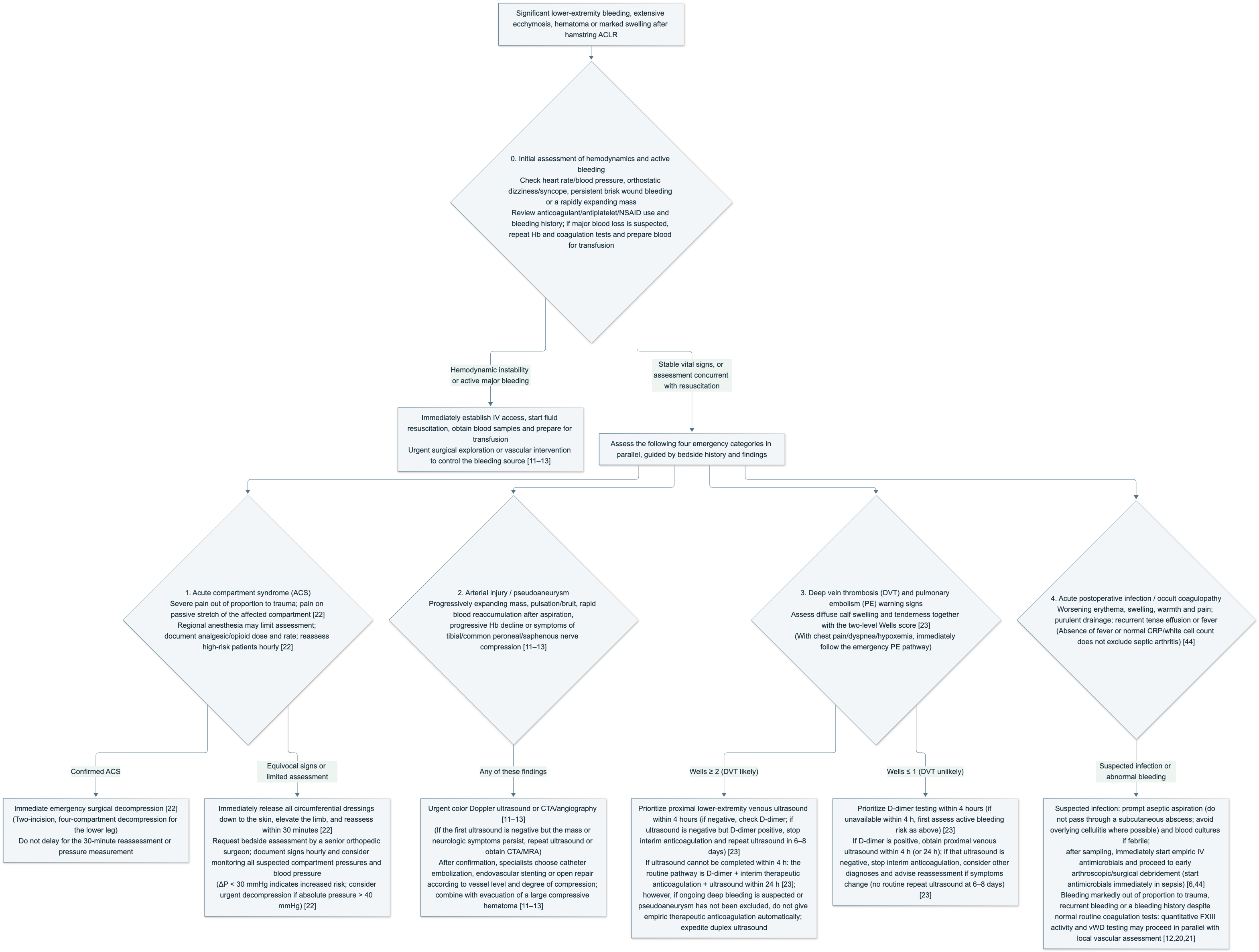
Figure 1. Flowchart for initial assessment and parallel emergency triage of significant lower-extremity bleeding, extensive ecchymosis, hematoma, or marked swelling after autologous hamstring ACLR (compiled from references [6,10–13,20–23,44]).
1. Initial Assessment of Systemic Hemodynamics and Bleeding Rate
Before focusing on the local limb, assess the systemic blood-loss burden:
-
Vital signs and tissue perfusion. Monitor heart rate, blood pressure, respiratory rate, and orthostatic symptoms (dizziness, visual darkening, cold sweat, or tachycardia on sitting or standing). Persistent active bleeding from the wound, a thigh or popliteal mass that expands visibly within hours, or hemodynamic instability warrants immediate intravenous access, fluid resuscitation, blood draw for type and crossmatch, and contact with emergency surgery or vascular interventional teams for hemostasis.
-
Correct interpretation of skin ecchymosis and early hemoglobin (Hb). The area and color depth of superficial subcutaneous ecchymosis reflect only the spread of blood into the superficial subcutaneous plane and its stage of degradation; they cannot quantify deep total blood loss. When deep massive or ongoing bleeding is suspected, serially repeat the complete blood count (Hb, hematocrit, platelets) and routine coagulation tests; early after acute blood loss, before interstitial fluid shift and intravenous fluid dilution are complete, a single early Hb value may not yet have fallen, and a single normal Hb cannot exclude acute massive bleeding.
-
Medication and bleeding-history review. Check perioperative prophylactic anticoagulants, antiplatelet agents, nonsteroidal anti-inflammatory drugs (NSAIDs), and personal or family bleeding history one by one (for example, prolonged bleeding after dental extraction, tonsillectomy, or trauma; childhood epistaxis lasting more than 30 minutes [11]; or known von Willebrand disease [12]).
2. Parallel Differentiation of Four Limb and Systemic Emergencies
(1) Acute compartment syndrome (ACS)
According to the British Orthopaedic Association Standards for Trauma (BOAST) revised in July 2025 [22], once high-tension swelling of the calf or thigh appears after ACLR, recognition and management should follow these standards:
-
Key clinical signs and analgesia monitoring requirements. Pain out of proportion to the degree of surgical trauma and pain on passive stretch of muscle in the affected compartment are the most important clinical findings [22]. BOAST requires documentation of pain severity and response to analgesia, doses and rates of analgesics (especially opioids), hourly assessment with written documentation of examination findings and the basis for management in patients at high risk of ACS, and clear assignment of postoperative ACS monitoring responsibility and guidance on limited assessment when regional anesthesia is used (clauses 1c, 3–6) [22]. On examination, sensory changes (numbness, pins and needles) may appear relatively early, whereas paralysis and skin pallor are late ischemic findings; the presence of a distal dorsalis pedis or posterior tibial pulse must never be used to exclude ACS or deep vascular injury [10,13].
-
Immediate release and 30-minute reassessment of suspected cases. Anyone with symptoms or signs suggestive of ACS must have all circumferential dressings, elastic bandages, and braces immediately released until the skin is fully exposed, with the limb elevated, and symptoms and signs reassessed within 30 minutes [22].
-
Immediate surgical decompression for confirmed cases and compartment pressure monitoring when uncertain. Once ACS is clinically confirmed, immediate surgical decompression must be performed; surgery must not be delayed for a 30-minute recheck or for pressure measurement [22]. If clinical signs are atypical or the patient cannot cooperate because of nerve block, a consultant orthopaedic surgeon with operative decision authority should assess at the bedside immediately, continue hourly documentation of signs, and consider pressure measurement of all suspicious compartments with simultaneous blood pressure: when the difference between diastolic pressure and compartment pressure (ΔP = diastolic pressure − compartment pressure) is < 30 mmHg, ACS risk is increased and the consultant decides between immediate surgical decompression and continued close monitoring; when absolute compartment pressure is > 40 mmHg, urgent surgical decompression should be considered [22]. For calf ACS, the standard procedure is two-incision, four-compartment complete decompression (in the rare cases with thigh involvement, decompress the affected thigh compartments); for late presentation or delayed diagnosis, given the very high risk of surgical complications, two senior specialists should jointly weigh operative versus nonoperative treatment, with concurrent assessment and protection of renal function [22].
-
Post-ACLR calf compartment hypertension cases and standard decompression. The 67-year-old man reported by Takahashi et al. developed severe calf pain, pallor, numbness, weakness, and medial blisters 12 hours after transtibial semitendinosus ACLR with ipsilateral medial and lateral meniscal repair without a tourniquet; CT showed tibial tunnel bleeding into the medial calf. Compartment pressures were not measured; ≤ 250 g of hematoma was evacuated through a medial longitudinal calf skin incision and medial fasciotomy, with primary skin closure and negative-pressure wound therapy (NPWT) [10]. This case confirms that severe tunnel bleeding and compartment hypertension can occur without a tourniquet, but the single medial fasciotomy with primary skin closure in this case report cannot replace the standard two-incision, four-compartment open decompression for calf ACS [10,22].
(2) Iatrogenic arterial pseudoaneurysm and peripheral nerve compression
Progressive swelling, a high-tension mass, rapid reaccumulation after aspiration, or accompanying neurologic symptoms at the distal medial thigh, around the tibial tunnel aperture, or in the popliteal fossa require focused evaluation for pseudoaneurysm [11–13]:
-
Deep pseudoaneurysms may lack surface pulsation, and distal pulses may be normal. When a pseudoaneurysm lies deep to sartorius or deep in the popliteal fossa and is wrapped in clot, surface palpation may detect no pulsation, and dorsalis pedis and posterior tibial pulses may also be normal [11,13]. The 19-year-old man reported by Ho et al. presented on day 6 with extensive ecchymosis from the posteromedial thigh to the calf and tenderness and swelling in the sartorius region, without a pulsatile mass, with normal distal pulses, negative D-dimer, and no thrombus on the first lower-extremity venous ultrasound; abnormality on CT followed by targeted color Doppler ultrasound and angiography finally confirmed a pseudoaneurysm of the saphenous branch of the descending genicular artery [11].
-
A single negative Doppler ultrasound cannot exclude popliteal artery pseudoaneurysm. Bonfim et al. reported two cases of delayed diagnosis of popliteal artery pseudoaneurysm after ACLR that caused severe tibial nerve injury [13]:
- Case 1 (34-year-old man). Immediate severe calf pain and paresthesia postoperatively, followed by loss of ankle and foot dorsiflexion and plantar flexion; after the first venous Doppler ultrasound excluded DVT, emergency calf fasciotomy was performed for suspected ACS. Because of worsening pain and complete paralysis, angiography on day 18 diagnosed popliteal artery pseudoaneurysm, treated with endovascular placement of seven covered stents; at about 3 months, nerve exploration showed continuity of the tibial nerve but a necrotic segment, which was resected and repaired with autologous sural nerve graft [13].
-
Case 2 (19-year-old woman). Persistent severe popliteal pain, a pulsatile mass, and skin blisters from day 2; the first Doppler ultrasound on day 4 was normal; diagnosis was established only on day 8 by magnetic resonance angiography (MRA; 45 × 52 mm partially thrombosed popliteal artery pseudoaneurysm) and repeat Doppler ultrasound, treated with endovascular stent repair (the text says “endovascular repair,” Fig. 3b “endovascular stent placement,” without stating whether covered) and surgical hematoma evacuation on day 13. At 12 months the common peroneal nerve recovered spontaneously while the tibial nerve remained completely paralyzed; exploration confirmed complete tibial nerve transection, repaired with sural nerve graft [13]. At 24-month follow-up after nerve repair, both patients had recovered ankle plantar-flexion strength to grade M4 but still had loss of toe flexion, plantar sensory impairment, and calf muscle atrophy [13].
-
Imaging reassessment requirements and limits of the original evidence. Bonfim et al. provided no objective intraoperative record excluding direct instrument injury to the tibial nerve during the original ACLR; the statements in their Discussion that “nerve compression for 3–4 days causes irreversible injury” and that CTA has “sensitivity 95.1%, specificity 98.7%” are also cited from other trauma literature [13]. These two courses nonetheless make clear that a first negative venous or arterial ultrasound never excludes deep pseudoaneurysm; once progressive neurologic symptoms or persistent deep severe pain appear postoperatively, timely repeat color Doppler ultrasound or direct CTA/MRA is mandatory [11,13].
-
Anatomic localization of peripheral nerve involvement. Bedside examination must strictly distinguish pure sensory from mixed-nerve involvement—traction or superficial hematoma compression of the saphenous or sural nerve causes only reduced sensation, numbness, or tingling in the medial calf/medial malleolus or posterolateral calf skin and does not weaken ankle or toe strength [4,7,11]; plantar numbness with ankle plantar-flexion or toe-flexion weakness (tibial nerve), or dorsal foot numbness with ankle dorsiflexion/toe-extension weakness or foot drop (common peroneal nerve), strongly suggests deep popliteal hematoma, pseudoaneurysm, or compartment hypertension involving a major nerve and mandates immediate emergency vascular imaging and surgical assessment [10,13,22].
(3) Lower-extremity deep vein thrombosis (DVT) and pulmonary embolism alerts
Calf swelling, tightness, and tenderness from harvest-site bleeding tracking into the calf closely resemble acute DVT. Following the National Institute for Health and Care Excellence (NICE) venous thromboembolism guideline (NG158) [23], bedside assessment and triage should follow these rules:
-
Use of the two-level Wells score. When history and examination still raise concern for DVT, use the two-level Wells score (≥ 2, DVT likely; ≤ 1, DVT unlikely) [23]. “Calf swelling at least 3 cm larger than the asymptomatic side” is only a single +1 item on the Wells score, not a specific confirmatory sign of DVT (deep hematoma can likewise enlarge circumference by >3 cm); postoperative ecchymosis or hematoma does not automatically earn the −2 deduction for “an alternative diagnosis at least as likely as DVT”—that deduction applies only when clinical examination confirms that bleeding adequately explains the present symptoms and is no less likely than DVT, noting that hematoma and DVT may coexist [23].
-
Pathways for Wells ≥ 2 and ≤ 1, and anticoagulation safety when active bleeding is suspected:
- Wells ≥ 2 (DVT likely; NG158 recommendations 1.1.3–1.1.7). Preferentially complete proximal lower-extremity venous ultrasound within 4 hours; if positive, manage as confirmed DVT; if negative, obtain D-dimer—if D-dimer is also negative, stop interim anticoagulation and consider other diagnoses (1.1.7); if the first proximal venous ultrasound is negative but D-dimer is positive, stop interim anticoagulation and repeat proximal lower-extremity venous ultrasound in 6–8 days (1.1.6) [23]. If proximal venous ultrasound cannot be completed within 4 hours, NG158 1.1.4 conventionally advises first obtaining D-dimer, giving interim therapeutic-dose anticoagulation, and completing proximal venous ultrasound within 24 hours [23].
-
Wells ≤ 1 (DVT unlikely; NG158 1.1.8–1.1.11). Wells ≤ 1 alone cannot exclude DVT; preferentially provide D-dimer within 4 hours (if D-dimer cannot be obtained within 4 hours, 1.1.8 conventionally advises interim therapeutic-dose anticoagulation while awaiting the result) [23]. If D-dimer is negative, stop interim anticoagulation and consider other diagnoses (1.1.9); if D-dimer is positive, complete proximal lower-extremity venous ultrasound within 4 hours per 1.1.10 (or give interim therapeutic-dose anticoagulation and complete ultrasound within 24 hours) [23]. Per NG158 1.1.11, for low-Wells patients with positive D-dimer but subsequently negative proximal venous ultrasound, manage directly per 1.1.7 (stop interim anticoagulation, consider other diagnoses, and advise the patient that DVT is currently unlikely and to seek reassessment if symptoms change); routine repeat ultrasound at 6–8 days, as in the high-Wells group, is not required [23].
-
Anticoagulation safety when ongoing bleeding or pseudoaneurysm is suspected after surgery. Clinical application must distinguish NG158 guideline text from individualized safety decisions in the setting of postoperative active bleeding. The NG158 Rationale and impact notes that interim therapeutic anticoagulation while awaiting tests rests mainly on committee experience and lacks direct clinical trial support; recommendation 1.3.7 on choice of anticoagulant requires weighing comorbidities, contraindications, and patient preference in principle [23]. On this basis, for patients with significant bleeding after ACLR, when deep active bleeding is still suspected, hematoma tension is progressively rising, or iatrogenic arterial pseudoaneurysm has not been excluded, do not mechanically apply empiric therapeutic-dose anticoagulation while awaiting ultrasound or D-dimer; instead urgently arrange duplex ultrasound of the lower extremity to examine deep veins, hematoma spaces, and arterial flow concurrently, with joint orthopaedic and vascular/thrombosis specialist decisions. Conversely, stable superficial subcutaneous ecchymosis alone without deep active bleeding is not a contraindication to anticoagulation.
-
Pulmonary embolism (PE) emergency triage. Chest pain, dyspnea, tachypnea, hemoptysis, or unexplained tachycardia and desaturation accompanying the swelling should immediately trigger the suspected acute PE emergency assessment and life-support pathway [23].
(4) Acute postoperative infection and occult coagulation factor deficiency
-
Recognition of early deep surgical-site infection and septic arthritis. Deep hematoma is a favorable culture medium for bacteria [5,6]. The Guideline for management of septic arthritis in native joints (SANJO 2023) was initiated by the European Bone and Joint Infection Society (EBJIS) and developed from systematic review; the ACLR infection chapter was completed with designated members of the European Society of Sports Traumatology, Knee Surgery and Arthroscopy (ESSKA); the publishing journal notes that the society guideline was not additionally peer-reviewed by the journal [44]. Progressive joint or donor-site erythema, swelling, warmth, and pain, purulent or cloudy wound drainage, rapidly recurrent tense joint effusion after aspiration, or systemic chills and fever should prompt assessment for infection at the first encounter; absence of fever or normal serum C-reactive protein (CRP) and white blood cell count never excludes septic arthritis. When clinically suspected, aspirate for sampling as soon as possible under strict asepsis (do not aspirate through a subcutaneous abscess, and avoid a path through overlying cellulitis when possible; Rec 8, D2), and obtain blood cultures concurrently in febrile patients or those with suspected bacteremia (Rec 7, 12) [44].
-
Evaluation for occult coagulation defects when routine coagulation tests are normal. When the severity of postoperative hemarthrosis or soft-tissue hematoma is clearly disproportionate to surgical trauma, bleeding recurs after aspiration or hemostasis, or there is a personal/family bleeding history with normal routine coagulation screening (PT/INR, aPTT, platelet count), factor XIII (FXIII) deficiency or von Willebrand disease (vWD) may be assessed in parallel while local vascular injury and drug factors are investigated; local iatrogenic vascular lesions and hereditary coagulopathy can coexist, as in the pseudoaneurysm patient with vWD reported by Barrio Piqueras et al. [12,20,21]. Tsujii et al. reported a 13-year-old female tennis player who underwent first arthroscopic medial plica resection for left medial synovial plica syndrome; the ACL and menisci were completely normal intraoperatively (not an ACLR). Two months later, after long-distance walking, she had recurrent left knee swelling; two aspirations each yielded 20 mL of bloody fluid, and MRI showed a hematoma and old clot in the suprapatellar pouch. Preoperative hematologic workup for unexplained hematoma before a second arthroscopy (resection of a complete-type suprapatellar plica septum and removal of old clot) showed normal PT, aPTT, bleeding time, platelet count, and von Willebrand factor, with only FXIII activity reduced to 54%. After the second arthroscopy she again developed severe hemarthrosis; on day 7 Hb fell from 13.5 g/dL to 10.3 g/dL (estimated blood loss about 800 mL); after FXIII concentrate on days 7, 8, and 9, FXIII activity rose to 98% on day 8 and 129% on day 9, bleeding stopped rapidly, and at 3 months FXIII activity had returned to baseline (57%) [20]. Per ARUP Laboratories testing standards (updated November 2023), FXIII maintains clot mechanical stability by catalyzing fibrin cross-linking; its deficiency does not prolong PT or aPTT at all, and traditional qualitative clot-solubility tests are sensitive only to extreme deficiency; therefore patients with normal routine coagulation but unexplained persistent or recurrent postoperative bleeding should have FXIII activity measured by a quantitative functional assay in conjunction with hematology (ARUP adult reference interval 69%–143%) [21]. Clinically, neither the 54% in the Tsujii case nor neurosurgical values they cite should be treated as a universal ACLR cut-off, still less should local vascular workup and surgical hemostasis be delayed while awaiting hematologic testing [20,21].
Table 1. Bedside Parallel Differential Points for Significant Lower-Extremity Swelling and Bleeding After Hamstring ACLR
| Clinical scenario | Bedside recognition clues | Common diagnostic traps | Confirmatory testing strategy | Emergency management principles |
|---|---|---|---|---|
| Acute compartment syndrome (ACS) [10,13,22] | Persistent pain out of proportion to surgical trauma; pain on passive stretch of the affected compartment [22]; board-like compartment tightness; early numbness/pins and needles may be present [10,13] | Presence of distal dorsalis pedis/posterior tibial pulse does not exclude ACS [10,13]; regional anesthesia impairs clinical assessment—document analgesic (especially opioid) dose and rate and reassess high-risk patients hourly [22] | Prefer clinical bedside diagnosis; when signs are uncertain or block limits assessment, measure pressures in all suspicious compartments and blood pressure (ΔP < 30 mmHg indicates increased risk; absolute pressure > 40 mmHg consider urgent decompression) [22] | For suspected cases, immediately release all circumferential dressings to skin, elevate the limb, reassess within 30 minutes; for confirmed cases, immediate surgical decompression (two-incision, four-compartment for the calf) [22] |
| Iatrogenic arterial pseudoaneurysm [11–13] | Progressively expanding mass at the medial thigh, proximal medial tibia, or popliteal fossa; rapid reaccumulation after aspiration; progressive Hb decline; or tibial/common peroneal motor-sensory deficit | Deep masses wrapped in clot may lack surface pulsation; distal pulses may be normal [11,13]; a single early Doppler ultrasound may be falsely negative [13] | Targeted color Doppler ultrasound; if the mass continues to enlarge or neurologic symptoms accompany a first negative ultrasound, must repeat ultrasound or urgently obtain CTA/MRA [11,13] | Specialist choice of catheter embolization, endovascular stent repair, or open repair according to vessel level and degree of compression; combine hematoma evacuation and nerve decompression/repair when high-tension large hematoma or nerve compression is present [11–13] |
| Lower-extremity deep vein thrombosis (DVT) [23] | Diffuse calf swelling and tightness; deep tenderness along deep veins; with chest pain/dyspnea, alert for pulmonary embolism (PE) | Postoperative ecchymosis does not automatically deduct 2 from Wells; calf circumference increase ≥ 3 cm is only one score item; negative venous ultrasound does not exclude arterial pseudoaneurysm | Two-level Wells: ≥ 2 prefer proximal venous ultrasound within 4 h (if ultrasound negative but D-dimer positive, repeat ultrasound at 6–8 days); ≤ 1 prefer D-dimer within 4 h, then ultrasound if positive (if that ultrasound is negative, manage per 1.1.7—no 6–8 day repeat) [23] | When deep active bleeding is still suspected or pseudoaneurysm has not been excluded, do not mechanically apply empiric therapeutic anticoagulation while awaiting tests; urgently arrange duplex ultrasound and multidisciplinary assessment |
| Acute postoperative infection (deep SSI / septic arthritis) [6,44] | Progressive local erythema, swelling, warmth, and pain; purulent/cloudy wound drainage; recurrent tense effusion; or chills and fever | Absence of fever or normal serum CRP and white cell count never excludes septic arthritis; hematoma and infection may coexist | Prompt aspiration under strict asepsis (not through a subcutaneous abscess; avoid overlying cellulitis path when possible); send for culture, Gram stain, white cell count with polymorphonuclear fraction, and crystal analysis; concurrent blood cultures if febrile/suspected bacteremia [44] | Unless sepsis/shock requires immediate antimicrobials, aspirate first then start empiric intravenous antibiotics per local resistance patterns and risk, and proceed promptly to arthroscopic/surgical debridement when early ACLR infection is suspected [6,44] |
| Gravity-dependent subcutaneous ecchymosis / stable donor-site hematoma [3,7,15] | Purple-red to yellow-green ecchymosis from posteromedial thigh to calf, medial malleolus, or foot; soft skin (or a deep, localized stable mass); no major-nerve motor deficit (occasional pure sensory numbness of saphenous/sural nerves) | Mistaking large gravity-dependent superficial ecchymosis for ongoing deep massive bleeding, or repeatedly blindly aspirating already organized solid clot with a large-bore needle | Bedside examination (ballotable patella / modified stroke test for effusion); musculoskeletal ultrasound of deep masses to distinguish liquid anechoic spaces from solid organized clot | Elevation, moderate elastic wrapping, and cold analgesia for superficial ecchymosis; high-tension liquid hematoma may undergo ultrasound-guided sterile aspiration after excluding vascular communication; organized compressive large hematomas undergo open or endoscopic evacuation |
3. Local Treatment and Specialist Management After Onset
After emergency triage is complete, clinical treatment depends on the anatomic compartment of bleeding, whether the hematoma is liquid or organized, local compressive symptoms, and whether vascular disease or infection is present.
1. Conservative Treatment of Extensive Gravity-Dependent Subcutaneous Ecchymosis and Stable Soft-Tissue Oozing
For patients with large purple-red or yellow-green ecchymoses of the posteromedial thigh, posteromedial calf, and ankle–foot region, but with soft local skin, no progressively enlarging high-tension mass, no major-nerve motor-sensory deficit, and stable systemic vital signs, conservative treatment is indicated:
-
Avoid blind aspiration and routine rescue antifibrinolytic medication. Diffusely distributed oozing in subcutaneous and intermuscular planes has no single concentrated fluid cavity; needle puncture cannot withdraw blood and instead increases the risk of carrying skin colonizers into a deep hematoma [36]. Published perioperative tranexamic acid (TXA) trials in ACLR [1,8,9,27,30,32,33] all used intraoperative or perioperative prophylactic dosing and did not test rescue dosing after ecchymosis or a stable hematoma had already formed; therefore rescue TXA is not routinely added for already stable postoperative ecchymosis or localized hematoma (if specialist assessment confirms ongoing active bleeding or a specific hyperfibrinolytic/coagulation disorder, manage per specialist hemostatic indications). Thoroughly explaining gravity tracking of blood along fascial planes into the calf and ankle and the natural color change from purple-red to yellow-green helps reduce patient anxiety.
-
Limb elevation, gentle elastic wrapping, and early cold analgesia. Elevate the limb at rest to promote venous and lymphatic return; after ACS, peripheral arterial insufficiency, and acute DVT have been clearly excluded, graduated elastic bandage wrapping within comfort limits may be used. Early postoperative ice or compression cryotherapy can reduce local pain and analgesic consumption [16,35,38], but a barrier should be used to prevent frostbite [16], and patients should be told that the main role of cold therapy is pain relief—it cannot be relied on to rapidly eliminate deep hematoma.
-
Careful review of concomitant antithrombotic and analgesic drugs. If the patient is taking anticoagulants, antiplatelet agents, or nonselective NSAIDs, the surgical team and the prescribing medical/anesthesia team should jointly weigh whether to hold or adjust therapy according to the primary antithrombotic indication, thrombotic risk, and whether bleeding has already stopped—never stop drugs indiscriminately without regard to indication. Stable superficial subcutaneous ecchymosis alone does not require interruption of necessary multimodal postoperative analgesia.
-
Warning symptoms requiring same-day urgent return. Outpatients or discharged patients must return the same day for reassessment if any of the following occurs: (1) thigh, popliteal, or calf swelling/mass enlarges or hardens rapidly within hours; (2) severe pain unrelieved by usual analgesics, or pain on passive calf stretch; (3) new plantar/dorsal foot numbness, or weakness of ankle dorsiflexion/plantar flexion or toe flexion–extension; (4) wound dehiscence, active bleeding, or cloudy/purulent drainage; (5) fever and chills, or sudden chest pain or dyspnea.
2. Aspiration Decompression of High-Tension Knee Hemarthrosis
Knee hemarthrosis and posteromedial thigh donor-site hematoma occupy different anatomic compartments and must be assessed separately—thigh subcutaneous ecchymosis does not mean there is necessarily a large intra-articular effusion, and joint aspiration will not evacuate a posterior thigh intermuscular hematoma [7,15,16].
When the patient has marked suprapatellar fullness, Coupens–Yates grade 3–4 high-tension hemarthrosis, and severe aching pain from capsular stretch, marked restriction of flexion–extension, and significant quadriceps AMI, knee aspiration and decompression should be performed via a superolateral approach under strict skin preparation and aseptic technique, followed by gentle elastic wrapping and early joint motion [8,9,14,19,27]. For example, the three patients with tense hemarthrosis on day 3 in the Bahl et al. study described in Section 1 all obtained symptom relief after sterile aspiration [14]; in the Michalski et al. case, early knee aspiration of 50 mL of blood (English abstract and text: within the first postoperative week; Polish text: the day after surgery) rapidly relieved the knee’s own aching pain, after which independent compressive symptoms of the deep calf intermuscular hematoma became apparent and received targeted ultrasound diagnosis [7]. If the joint rapidly refills with fresh blood shortly after aspiration decompression, do not simply aspirate repeatedly—immediately obtain color Doppler ultrasound/CTA and coagulation testing to evaluate for periarticular arterial branch pseudoaneurysm or coagulation factor deficiency [12,20,21].
3. Staged Intervention for Encapsulated Hematoma of the Thigh Donor Site and Calf Intermuscular Space
When harvest-tract bleeding accumulates deep in the posteromedial thigh or tracks into the gastrocnemius–soleus interval as a localized hematoma, first determine whether bleeding is still ongoing: if active bleeding is present, the priority is to control the source; before needle aspiration of any deep hematoma, color Doppler ultrasound (CTA if needed) must first confirm a liquefied cavity and exclude an arterially communicating pseudoaneurysm—never advance a needle blindly merely because gray-scale ultrasound shows a hypoechoic dark area [7,11–13,36]. After vascular lesions are excluded, because the speed of organization varies individually, specific intervention depends on the actual hematoma morphology on ultrasound or MRI (liquid cavity versus solid organized clot) and the trend of local compressive symptoms, guided by real-time imaging morphology rather than fixed week cut-offs [7,36]:
-
Localized hematoma of mild tension without neurovascular compression (conservative observation and serial reassessment). If the deep hematoma is small, local skin has no tight blisters, pain is not progressively worsening, and there are no distal neurologic symptoms, prefer limb elevation, gentle elastic wrapping, and scheduled review, confirming gradual softening and shrinkage by serial monitoring of circumference, local tension, and neurologic status.
-
Liquid-phase hematoma causing marked aching pain or compressive symptoms (ultrasound-guided sterile aspiration). If deep hematoma tension is high, causing significant aching pain or impeding knee and ankle motion, and Doppler ultrasound confirms a liquefied cavity without flow signal, aspirate under real-time ultrasound guidance and strict asepsis, then apply moderate compression wrapping to close dead space and relieve local tension [36]. Per the GOTS muscle injury expert consensus, no clinical trial has yet directly shown that aspirating hematoma shortens tissue healing time or reduces reinjury rate; the main clinical value of aspiration is rapid relief of local high tension and pain. Meanwhile, the “maximum tolerable pressure compression” and fixed day-2 and day-5 ultrasound recheck patterns that GOTS proposed for acute muscle tears on the field cannot be applied directly after ACLR; postoperative compression must also respect wound healing and compartment safety [36].
-
Already organized solid clot and recurrent/compressive deep large hematoma (avoid repeated large-bore blind aspiration; timely open or ultrasound-assisted endoscopic evacuation). When ultrasound or MRI shows the hematoma cavity filled with dense fibrin clot and organized tissue, needle aspiration cannot empty the clot [7,36]. In the 48-year-old man reported by Michalski et al. after autologous semitendinosus–gracilis ACLR, ultrasound on day 14 showed a large hematoma between gastrocnemius and soleus; ultrasound-guided 18G large-bore aspiration yielded only a very small amount of organized blood; at week 3 an attempt to evacuate 250 mL of organized hematoma openly through the original tibial harvest-incision scar was followed by recurrence within 7 days. At week 5 they performed ultrasound-assisted endoscopic hematoma evacuation: ultrasound marked the superior and inferior poles pre- and intraoperatively; dual posteromedial calf portals were established; organized clot was cleared under direct vision, fibrous adhesions released, and a suction drain placed; intraoperatively, saline was drip-infused by gravity alone to maintain the view (no pressurized arthroscopic pump) to prevent extravasation into intermuscular planes inducing iatrogenic compartment syndrome; postoperative calf swelling, pain, and nerve-irritation symptoms resolved completely [7].
This article is an uncontrolled single-case report; its abstract’s portrayal of aspiration as the sole “gold standard” and assertion that endoscopy is generally superior to open surgery lack comparative evidence. Combined with the treatment course, however, three practical points can be drawn for managing such deep organized hematomas [7]:
- Do not repeatedly aspirate with a large-bore needle when ultrasound confirms already organized solid clot, lest deep infection risk be needlessly increased [7,36];
- If open evacuation is performed for persistently compressive or recurrent organized large hematoma, the approach must provide adequate exposure of the corresponding hematoma cavity; squeezing distally through a proximal original harvest mini-incision alone may fail to empty a deep residual calf cavity thoroughly [7];
- Ultrasound-assisted soft-tissue endoscopic evacuation is a minimally invasive option for deep recurrent organized hematoma; gravity drip infusion without a high-pressure pump helps prevent extravasation-induced compartment hypertension [7].
4. Specialist Management of Confirmed Iatrogenic Arterial Pseudoaneurysm and Concurrent Deep Infection
- Iatrogenic arterial pseudoaneurysm. Vascular surgery or interventional radiology selects an individualized plan according to the level of the affected artery, collateral circulation, hematoma volume and tension, and whether nerve compression or infection is present [11–13]:
- Periarticular arterial branch lesions (for example, saphenous branch of the descending genicular artery, articular branch of the inferior medial genicular artery). Options include catheter embolization of the diseased vessel under superselective angiography with micro-coils (as in the Ho et al. case [11]) or liquid embolic agent (SquidPeri 18, as in the Barrio Piqueras et al. case [12]), or open ligation/repair; case reports only show that these embolization techniques were feasible in the corresponding cases and cannot establish a single preferred procedure or specific embolic material [11,12]. The absolute statement by Ho et al. that collateral circulation guarantees “no risk of any skin or tissue necrosis” after embolization should not be accepted [11]; local skin perfusion must still be observed postoperatively. If a huge high-tension hematoma has already formed beside the pseudoaneurysm (about 600 mL in the Ho et al. case) and compresses the saphenous nerve or surrounding muscle, surgical evacuation of hematoma for rapid decompression should be combined after interventional embolization interrupts arterial inflow [11].
-
Popliteal artery trunk pseudoaneurysm or progressive tibial/common peroneal nerve injury. As in the two popliteal artery pseudoaneurysms reported by Bonfim et al., vascular surgery may perform endovascular stent repair according to lesion morphology (case 1 received seven covered stents; case 2 is described as endovascular stent repair without stating whether covered) or open vascular repair; when a huge hematoma compresses nerve or soft tissue, timely surgical hematoma evacuation is needed (as in Bonfim case 2, open hematoma drainage after stenting), with close postoperative follow-up of tibial and common peroneal nerve function; if paralysis fails to recover, timely peripheral nerve exploration, neurolysis, or autologous sural nerve grafting is required [13].
-
Deep surgical-site infection (SSI) or septic arthritis secondary to hematoma. Following SANJO 2023 [44] and principles of post-ACLR infection management [6], once deep infection or septic arthritis secondary to hematoma is suspected:
- Aspirate and obtain blood cultures first, then start empiric antimicrobials immediately (except in sepsis). Promptly aspirate the joint cavity or deep fluid space under strict asepsis; do not aspirate through a subcutaneous abscess, and avoid a path through overlying cellulitis when possible (SANJO Rec 8, D2); send specimens for aerobic/anaerobic culture, Gram stain, white cell count with polymorphonuclear fraction, and crystal analysis, and obtain blood cultures concurrently if febrile or bacteremia/sepsis is suspected (Rec 7, 12) [44]. After sampling, immediately start empiric intravenous antimicrobials according to local pathogen epidemiology and resistance patterns, Gram stain results, and patient risk factors for resistant organisms (for example, methicillin-resistant Staphylococcus aureus [MRSA] or multidrug-resistant Gram-negative bacilli), covering S. aureus, coagulase-negative staphylococci, and streptococci/Gram-negative bacilli among potential pathogens (Rec 12–13; culture-positive cases in the Sever cohort grew S. aureus or coagulase-negative staphylococci such as S. epidermidis [6]; if the patient already has sepsis or septic shock at presentation, give drugs immediately with fluid resuscitation and do not delay antimicrobials for aspiration, Rec 12) [6,44].
- Early surgical/arthroscopic debridement and drainage. SANJO recommends arthroscopic debridement as the principal operative option for post-ACLR infection (Rec 19, C2), assessing the graft by arthroscopic exploration and debridement to control infection early and promote recovery (B1); for cases with acute symptoms or in the early postoperative period, once infection is clinically suspected, proceed to arthroscopic debridement promptly even if microbiology results are not yet available (Rec 19, B1); if concurrent extra-articular deep donor-site hematoma infection is present, perform open debridement and drainage concurrently [6], then adjust to targeted antimicrobials once culture and susceptibility results are available [6,44].
4. Pausing, Preserving, and Advancing Postoperative Rehabilitation According to Response
1. Current Evidence and the Principle of “Extension Activation—Donor-Site Posterior-Chain Separated Management”
Current ACLR rehabilitation clinical practice guidelines and RCTs mainly address routine postoperative populations without major bleeding complications; among published clinical research, no prospective clinical trial has specifically tested the optimal number of rest days, fixed waiting weeks, or fixed percentage load increments for patients with substantial donor-site bleeding or hematoma after autologous hamstring ACLR [15,16,36,37]. Therefore, load reduction, preservation, and progression for these patients must be synthesized from criterion-based rehabilitation principles for routine ACLR (for example, Adams et al. [15] and the Aspetar guideline [16]), muscle hematoma management consensus (GOTS [36]), and the actual tissue response of the operated limb; fixed rest days or uniform load doses for this hematoma population have not been validated.
When planning rehabilitation clinically, the key is to manage knee extension/quadriceps activation separately from posteromedial thigh hamstring donor-site load, following these basic principles:
-
Load pause and safe preservation during emergency triage and active bleeding. Aspetar (Box 3) supports early active motion, isometric quadriceps training, and early progressive weight-bearing after routine ACLR [16]; but during emergency triage when the hematoma is progressively enlarging, local pain is severe, or ACS, pseudoaneurysm, acute DVT, and deep infection have not been excluded, training loads that raise compartment/venous pressure or traction the bleeding cavity must be paused, including active/resisted knee flexion, posterior-chain stretching, prolonged dependent standing, high-load weight-bearing walking, and blood-flow restriction (BFR) training [16,22,44]. Unnecessary prolonged immobilization should still be avoided lest muscle atrophy and venous stasis worsen, but when emergency assessment is incomplete, or active bleeding, suspected DVT, or suspected septic arthritis/deep infection persists, full bedside knee training must not be restored solely because ACS and unstable vascular lesions have been excluded. The treatment team should decide the scope of bedside activity according to whether active bleeding is controlled, progress of DVT workup and anticoagulation, and infection control: with concurrent septic arthritis, while drains remain, the wound is open, and infection is uncontrolled, maintain only distal ankle pumps and other activities that do not involve the affected knee [44]; when bleeding is controlled, acute infection of the affected joint is excluded, and thrombosis management allows, pain-free low-load bedside activities such as distal ankle pumps, gentle patellar mobilization, and isometric quadriceps contraction in extension may be preserved [15,16,23,44].
-
Superficial subcutaneous ecchymosis alone does not limit routine rehabilitation progression. Intra-articular hemarthrosis, deep encapsulated hematoma, and superficial gravity-dependent subcutaneous ecchymosis must be distinguished. The area and color of subcutaneous ecchymosis are not indicators that limit rehabilitation; as long as the skin is soft, there is no deep tight hematoma mass or severe tenderness, and joint effusion is stable, even large purple-blue or yellow-green ecchymoses from calf to ankle allow normal progression of full passive/active extension (0°), isometric quadriceps contraction, straight-leg raise, and crutch-assisted weight-bearing gait within tolerance by usual standards [15,16].
-
Accurate grasp of biomechanical stretch relationships of lower-extremity biarticular muscles. The hamstrings (semitendinosus, semimembranosus, long head of biceps femoris) and gracilis cross the hip and knee (gracilis lies medially in the thigh). Biomechanically, hip flexion with simultaneous knee extension (for example, high-angle supine straight-leg raise, sitting or standing forward bend with knee extension) directly lengthens the hamstrings and the harvest-tract wound; isolated passive knee flexion (with the hip neutral or slightly flexed) does not lengthen the hamstrings. Therefore, in the acute phase of donor-site hematoma or high-tension hemarthrosis, both high-angle hip-flexion–knee-extension stretch of the posterior harvest cavity and forced deep painful knee flexion should be avoided lest capsular or posterior soft-tissue aching pain worsen [15,16].
2. Timing of Hamstring Donor-Site Load Restoration and Appraisal of Two Literature Protocols
On when to begin loading after autologous hamstring harvest, the literature contains two protocols with markedly different timelines; neither should be applied mechanically in clinic:
-
The 12-week delay rule of Adams et al. (2012) [15]. Considering muscle belly retraction, donor-site pain, and soft-tissue healing after autologous hamstring harvest, that guideline’s body and appendix precautions specify no resisted hamstring strengthening for the first 12 postoperative weeks, with gradual resistance thereafter by a soreness rule [15]. This is conservative experiential protection within a specific rehabilitation system and has not been validated by controlled trials as the sole safe starting point.
-
Ricupito et al. (2025): early open kinetic chain (OKC) training from week 3, with no restart before week 8 after a safety stop [37]. This single-group retrospective study (n=13, all young active patients after autologous semitendinosus–gracilis ACLR) began twice-weekly seated single-leg 90° and 60° isometric knee flexion and 30° long-lever bridges from week 3 (5 sets × 45 s, pain kept NPRS < 3/10), progressing to seated leg-curl progressive isotonic resistance from week 6; the authors set three OKC safety red lines (pain > 4/10 during training or within 24 hours; visual/palpatory or ultrasound discovery of posterior thigh/donor-site hematoma; signs of hamstring strain), requiring OKC to stop if triggered until completely pain-free and not earlier than week 8 for restart [37]. However, enrollment strictly excluded any clinically meaningful complication in the first 3 postoperative weeks and required no effusion at enrollment (stroke test below Trace) and resting pain ≤ 3/10; across 234 training sessions in 13 patients, actual hematoma occurrence was 0, and the single recorded pain of 4.4/10 (incidence 0.4%), although above the method’s stop threshold, was noted in the Results as not actually interrupting that session [37]. Therefore, neither can these 13 strictly screened hematoma-free cases support the inference that “patients who have already had a large donor-site hematoma are equally safe with resisted knee flexion at week 3,” nor should this center’s “8 weeks” or Adams’s “12 weeks” be treated as a fixed interval that every hematoma patient must wait until 100% hematoma resolution before gentle posterior-chain activation [15,37].
Donor-site load restoration ladder and joint-motion conditions after debridement:
- Posterior-chain load progression after donor-site or calf hematoma stabilizes. When serial observation confirms that donor-site or calf bleeding has completely stopped, deep hematoma tension has clearly fallen with softening and shrinkage, local resting and palpation tenderness has markedly lessened, effusion is controlled, and the patient has good pain-free extension control, posterior-chain activity may be cautiously resumed through the following stages (if concurrent meniscal repair, cartilage repair, or multiligament surgery is present, angles and weight-bearing still follow the concurrent surgery’s protection requirements) [15,16,37]:
- Stage 1 (unresisted ROM and submaximal isometric activation). Pain-free active-assisted knee flexion ROM (for example, supine pain-free heel slides), then trial of low-intensity, short-lever submaximal isometric contraction and bilateral closed-chain bridges at pain-free angles.
-
Stage 2 (single-variable progressive low-load closed-/open-chain training). If the donor site has no stabbing or cramping pain on the training day and the next morning, hematoma tension and limb circumference do not rebound, and effusion does not increase, progress gradually to single-leg bridges and light-load seated/prone isotonic knee flexion; increase only one training variable slightly each time (hold time, repetitions, or load). If donor-site pain worsens, hematoma tension rises, or circumference rebounds during training or the next day, immediately return to the prior tolerated level and reassess [15,16,37].
-
Distinguishing rehabilitation start after sterile organized hematoma evacuation versus infection debridement:
- After sterile organized large hematoma evacuation. Once no active rebleeding and no risk of wound dehiscence are confirmed, there is no need to wait for complete absorption and closure of the deep residual cavity; under surgical team guidance, bedside pain-free ankle pumps, passive extension (0°), gentle patellar mobilization, and isometric quadriceps contraction may begin, with progressive restoration of weight-bearing and closed-chain training as drains are removed and the wound heals [7,15,16].
- After debridement for concurrent septic arthritis or deep infection. Strictly distinguish distal activity that does not involve the affected knee from activity of the affected knee—while drains remain or the wound is not yet closed, the patient may perform bedside pain-free ankle pumps that do not move the affected knee to reduce venous stasis; motion of the affected knee should follow SANJO 2023 recommendation 15 and begin as early as possible after infection is controlled, drains are removed, and the surgical wound is closed (D2) [44], then progressively restore weight-bearing and strengthening according to tissue response [15,16,44].
3. Bedside Daily Monitoring Tools: Modified Stroke Test, Local Signs, and Soreness Rule
Daily rehabilitation advance or retreat rests on the following bedside monitoring indices (the soreness rule in Adams et al. Table 3 was originally cited from Fees et al. 1998 on upper-extremity strength training [15] and, for this patient group, must be combined with donor-site hematoma tension and circumference monitoring):
- Modified stroke test for knee effusion (Adams 2012 Table 2 / Sturgill 2009) [15]. The examiner first gently strokes 2–3 times from the medial joint line toward the proximal suprapatellar pouch to empty the medial recess, then with the other hand strokes down the distal lateral thigh from the suprapatellar pouch and observes the fluid wave in the medial parapatellar gutter:
- Grade 0 (Zero): no medial fluid wave on the downstroke;
- Trace: a small medial fluid wave on the downstroke;
- 1+: a larger bulging medial fluid wave on the downstroke;
- 2+: after the upstroke, effusion spontaneously returns to the medial side of the knee without a downstroke (effusion spontaneously returns to medial side after upstroke, no downstroke necessary);
- 3+: effusion volume so large that fluid cannot be milled away from the medial side of the knee [15].
Adams et al. use modified stroke test grade 0 or Trace as one criterion for transition to high-load stages such as running and jumping in their standard ACLR progression [15], but that grade cut-off has not been validated as a uniform load-reduction threshold in patients with postoperative hematoma; clinical monitoring of these patients should judge whether to reduce training load by whether the modified stroke test grade has risen relative to the patient’s baseline, combined with joint and donor-site pain, posterior thigh/calf hematoma tension, circumference change, and functional response [15,16].
-
Local donor-site and calf sign monitoring. Daily measure circumference at fixed anatomic landmarks (for example, a fixed distance proximal to the superior pole of the patella and the maximal calf circumference), and palpate posteromedial thigh and gastrocnemius tension, tenderness, skin temperature, and aspiration/debridement incision status.
-
In-session and next-morning symptom response rule [15,16]. If soreness appears during warm-up and does not resolve, or resolves after warm-up but reappears during training, or joint/donor-site pain that is not delayed-onset muscle soreness appears the next morning with circumference rebound, pause the current progression and step down one load level; advance steadily only when there is no adverse response on the day and the next morning [15].
4. Evidence Appraisal and Contraindications for Adjunctive Physical and Rehabilitation Interventions
- Cryotherapy and compression cryotherapy (for early analgesia; no evidence of promoting large hematoma absorption). Aspetar notes that cryotherapy helps reduce pain in the first postoperative week, lower analgesic need, and improve satisfaction, but swelling reduction is inconsistent (overall evidence certainty very low) [16]. The systematic review by Daszkiewicz et al. (2026) included 9 controlled trials (509 ACLR patients in total; the review’s appendix quality assessment reports that 1 trial allocated by odd/even last digit of the social security number, so not all included studies can be treated as strictly randomized): 4 trials reported cryotherapy reduced early pain, 2 improved early ROM, and the 4 trials assessing blood loss and wound drain output (270 patients) all found no significant difference; GRADE certainty was very low for all outcomes [35]. The small pilot of Dambros et al. (analyzed ice group 10 vs control 9) likewise observed only the effect of two 20-minute ice applications on day 1 on pain and ROM and did not measure blood loss or swelling [34].
The randomized trial of Yang et al. (device group 10°C/30 mmHg and ice-pack group, 28 each; 52 autologous hamstring, 4 allograft) showed lower VAS pain on day 4 in the device group (2.1 ± 1.4 vs 3.3 ± 1.3, P = 0.001), but no difference in pain or knee circumference on days 7 and 14; in their Table 5, separate comparison of 48-hour drain output (28 each: 148.2 ± 102.8 mL vs 171.3 ± 64.8 mL, P = 0.324) and of day-6 MRI joint effusion volume (21 vs 22 completing MRI: 47.1 ± 11.9 mL vs 51.8 ± 16.0 mL, P = 0.280) both showed no significant difference; only the composite “48-hour drain output + day-6 MRI joint effusion” in the 43-patient MRI subgroup differed (175.2 ± 83.4 mL vs 239.7 ± 78.8 mL, P = 0.015), and that composite reflects only joint fluid, not absorption of thigh donor-site hematoma [38]. The staged cohort of Moussa et al. (2023) (dynamic intermittent compression cryotherapy plus intravenous nefopam, 338, vs static compression cryotherapy plus oral nefopam, 338) changed both cryotherapy mode and analgesic route simultaneously, and baseline rates of concurrent lateral extra-articular tenodesis differed significantly (40.2% vs 21.0%, P < 0.001), so it cannot prove that compression cryotherapy eliminates hematoma [39]. Clinically, therefore, cryotherapy is positioned as a safe early analgesic adjunct.
- Neuromuscular electrical stimulation (NMES; supported as an adjunct for quadriceps activation; exclude the retracted paper claiming DVT prevention and swelling reduction). Aspetar (Box 2) supports early NMES to help overcome AMI and activate the quadriceps [16]. The double-blind sham-controlled trial of Toth et al. (25 randomized, 21 completed; at 3 weeks, muscle biopsy fiber cross-sectional area NMES 11 vs sham 9, single-fiber contractile force 10 vs 9; at 6 months, whole-muscle cross-sectional area and strength 9 vs 7) showed that early NMES (5 days/week, 1 hour/day, through week 3) attenuated MHC II fiber atrophy in vastus lateralis at 3 weeks and preserved MHC I fiber contractile force [40]. Almost all subjects received BPTB grafts (Table 1: autologous hamstring only 1 in sham, 0 in NMES); whole-muscle strength at 6 months did not differ between groups; in Supplementary Table 1, the operated-to-uninjured quadriceps cross-sectional area ratio at 6 months was 72.2 ± 2.4% vs 82.8 ± 3.6% (mean ± SEM, P = 0.023; at 3 weeks 70.1 ± 1.9% vs 73.8 ± 3.0%, P = 0.273) [40]. Clinically, NMES may be used to assist neuromuscular quadriceps recruitment, but not as a means to eliminate hematoma or guarantee long-term whole-muscle hypertrophy.
In addition, Xiong et al. (2022) claimed that NMES after ACLR lowers D-dimer, reduces lower-extremity swelling, and prevents DVT [41]; that article was formally retracted on 30 August 2023 (PMID 37680554). The retraction notice states that publisher investigation found signs of systematic manipulation of the publication process, and that at least one of the human-subjects reporting requirements—institutional review board (IRB) or equivalent ethics approval, informed consent of patients/subjects to participate, or (where applicable) consent to publish personal details—was not met; the notice also states that whether the authors knew of or participated in the publication-process manipulation was not investigated [42]. Combined with the original section 3.2’s contradictory claims within the same paragraph that day-4 VAS, D-dimer, and circumference both “did not differ significantly” and were “significantly lower” [41], clinical citation of this retracted study to support NMES for thrombosis prevention or swelling reduction is forbidden.
- Contraindications and hold conditions for trigger-point dry needling and blood-flow restriction (BFR) training:
- Trigger-point dry needling. In the randomized trial of single-session vastus medialis dry needling in the subacute phase after ACLR (postoperative days 7–21) by Velázquez-Saornil et al. (dry needling plus rehabilitation 22 vs rehabilitation alone 22), the dry-needling group had 3 cases (13.6%, 3/22) of local bleeding adverse events with area > 4 cm² (1 withdrew for this reason; control 0/22, P = 0.073), and immediate post-needling pain was greater [43]. Aspetar (Box 2) accordingly does not recommend vastus medialis dry needling in the very early postoperative period [16]. For early postoperative patients and those with significant bleeding or deep hematoma not yet stable, invasive needling such as dry needling should be deferred [16,43].
- Blood-flow restriction (BFR) training. Aspetar (Box 2) explicitly lists extensive swelling and vascular disease as contraindications to avoid for low-load BFR [16]. Until significant bleeding, marked swelling, or vascular disease of the operated limb has been fully excluded, BFR cuff training is prohibited.
Table 2. Brief Dynamic Adjustment of Postoperative Local Management and Rehabilitation Training by Clinical State
| Clinical state category | Bedside findings and confirmation conditions | Local medical and surgical management focus | Rehabilitation loads to pause and contraindicated procedures | Training to preserve and restore progressively by response |
|---|---|---|---|---|
| Scenario A: Emergency triage / active bleeding, suspected ACS, pseudoaneurysm, or pending DVT/infection workup | Severe pain, pain on passive stretch, or high compartment tension [22]; progressively enlarging mass or major-nerve motor-sensory deficit [11–13]; diffuse calf tenderness or erythema/fever [23,44] | Immediate surgical decompression if ACS confirmed (for suspected cases, release dressings to skin, elevate, reassess in 30 minutes, and measure pressures) [22]; urgent duplex ultrasound/CTA or aspiration culture [11,13,23,44] | Pause active/resisted knee flexion, posterior-chain stretching, prolonged dependent standing, high-load weight-bearing, BFR, and dry needling [16,22,43] | Avoid unnecessary prolonged immobilization, but while emergency assessment is incomplete, active bleeding persists, or DVT/infection is suspected, do not restore all bedside training solely because ACS and unstable vascular lesions are excluded; the treatment team decides which pain-free low-load bedside activities may be preserved according to bleeding control, thrombosis anticoagulation status, and infection management (for example, distal ankle pumps, or gentle patellar mobilization and isometric quadriceps in extension when bleeding is controlled, acute infection of the affected joint is excluded, and thrombosis management allows) [15,16,23,44]; once the emergency is resolved and bleeding stable, transition to scenarios B–D |
| Scenario B: Extensive gravity-dependent subcutaneous ecchymosis (no deep high-tension hematoma mass) | Purple-red to yellow-green ecchymosis from posteromedial thigh to calf/ankle–foot; soft skin, no deep tight mass; motor nerves normal; no high-tension hemarthrosis, effusion stable [15,16] | No aspiration, no routine added hemostatic drugs; explain gravity-tracking natural history; elevate limb, elastic wrapping within comfort, early safe cold analgesia [16,35] | Defer vastus medialis dry needling in the very early postoperative period; defer BFR until marked swelling resolves [16,43] | Subcutaneous ecchymosis area does not limit routine rehabilitation: progress normally to 0° full extension, isometric quadriceps, straight-leg raise, NMES-assisted activation, and progressive weight-bearing gait within tolerance [15,16,40] |
| Scenario C: Medium-to-large localized donor-site/intermuscular hematoma or high-tension hemarthrosis (no ischemic emergency) | Localized hematoma mass in posteromedial thigh or gastrocnemius–soleus interval [7]; or Coupens–Yates 3–4 high-tension hemarthrosis with marked AMI [8,14,15] | Strict sterile aspiration decompression for Coupens–Yates 3–4 high-tension hemarthrosis with significant aching pain, marked flexion–extension restriction, or significant quadriceps AMI [8,9,14,19,27]; first exclude vascular communication of deep hematoma by Doppler ultrasound; liquid high-tension cavities causing marked aching pain or impeding motion may undergo ultrasound-guided sterile aspiration; do not repeatedly blindly aspirate solid organized clot [7,36] | Defer isolated resisted knee flexion, high-angle hip-flexion–knee-extension stretch, forced painful deep flexion, dry needling, and BFR [15,16,37,43] | Separate progression of extension and flexion loads: continue pain-free extension, isometric quadriceps, and controlled weight-bearing; after hematoma tension falls and tenderness eases, restore posterior-chain load by the ladder “pain-free heel slides → submaximal isometric/bilateral bridges → progressive resistance,” stepping down if next-day effusion grade rises above baseline or hematoma tension rebounds [15,16,37] |
| Scenario D: After evacuation of refractory/recurrent organized large hematoma or after infection debridement | After open or ultrasound-assisted endoscopic (gravity drip) evacuation of organized large hematoma [7]; or after debridement/drainage and antimicrobials for early septic arthritis/deep SSI [6,44] | Protect the surgical wound and drains; monitor for reaccumulation, skin perfusion, and infection-control indices (temperature, CRP, drain-fluid character) [6,7,44] | While the wound is open, drains remain, or infection is uncontrolled, defer forceful flexion–extension of the affected knee, direct pressure on the incision, high-load resistance, and hydrotherapy/invasive procedures [44] | Distinguish sterile hematoma evacuation from infection debridement: after sterile hematoma evacuation, once no rebleeding is confirmed, begin bedside pain-free ankle pumps, passive extension (0°), patellar mobilization, and isometric quadriceps activation [7,15,16]; after septic arthritis debridement, while drains remain keep only bedside distal activity that does not involve the affected knee (for example, ankle pumps); begin affected-knee motion as early as possible after infection is controlled, drains are removed, and the surgical wound is closed (SANJO Rec 15, D2) [44], then progressively restore weight-bearing and closed-chain training [15,16,44] |
5. Perioperative Preventive Measures and Evidence Appraisal
Prevention of significant bleeding and hematoma after autologous hamstring ACLR involves intraoperative harvest hemostasis, drain-placement strategy, and perioperative antifibrinolytic drug use. The great majority of existing perioperative trials measured only 24- to 48-hour joint drain output or early hemarthrosis grade and did not directly count the incidence of large posteromedial thigh hematoma or extensive calf ecchymosis, and some studies have design confounding or data errors (Table 3).
1. Harvest Anatomic Separation and Intraoperative Hemostasis
-
Separate accessory tendon connections first; avoid blind advancement of scissors. Before advancing the stripper, identify and divide the accessory tendon connections (fascial expansions) of semitendinosus and gracilis to the medial gastrocnemius fascia under direct vision or by tendon traction combined with finger palpation, to prevent premature tendon amputation or tearing of larger muscle-belly vessels [4,17,18]. Opinion differs on dry endoscopic-assisted harvest, but even with endoscopy injury to the infrapatellar branch of the saphenous nerve (IPBSN) cannot be completely avoided [17,18]. When separating accessory tendons, never blindly push open scissors deep to sartorius, lest the saphenous branch of the descending genicular artery be injured [4,11].
-
Local donor-site compression and direct-vision hemostasis versus routine additional intra-articular radiofrequency coagulation. In a technical guide, Familiari et al. propose packing the harvest tract after harvest with a slender Raytec gauze strip soaked in 10 mL of TXA solution (500 mg/5 mL) for compression hemostasis, and coagulating small vessels around the donor site and tibial tunnel [4]; that article is uncontrolled expert experience and reports no hematoma incidence data [4]. In a randomized trial of 60 single-bundle hamstring ACLR cases, Çimen et al. compared radiofrequency probe coagulation of bleeding points in the intercondylar notch (middle genicular artery) and Hoffa fat pad after tourniquet release at the end of arthroscopy (n=30) with no additional intra-articular coagulation (n=30; neither group drained) and found that routinely adding arthroscopic intra-articular coagulation did not reduce Coupens–Yates hemarthrosis grade from day 1 to day 45, aspiration rate (2/30 [6.7%] vs 4/30 [13.3%], P = 0.389), VAS pain, or thigh circumference, and instead prolonged anesthesia time (79.5 ± 20.8 min vs 63.4 ± 16.0 min, P = 0.001; mean day-1 tramadol in their Table 4 of 253.3/276.7 mg also exceeded the routine daily dose of 200 mg stated in Methods) [19]. The Çimen trial assessed only intra-articular soft tissue and did not address the extra-articular hamstring harvest tract at all; therefore this negative result only indicates that routinely prolonging radiofrequency burn time inside the joint is unnecessary and in no way negates the need for direct-vision hemostasis of visible active bleeding points at the harvest incision and tibial tunnel aperture [4,19].
2. Tourniquet Use and Drain Strategy (Strict Distinction Between Donor-Site and Joint-Cavity Drains)
-
Effect of tourniquet use. In a historical controlled cohort, Nagashima et al. compared an inflated tourniquet during the arthroscopic stage (T+ group, 55 knees/55 patients, 2017–2018, gravity irrigation) with no tourniquet throughout (T− group, 77 knees/76 patients, 2018–2021, 40–80 mmHg pump irrigation; neither group inflated a tourniquet during harvest; both included 10 revisions and a 24-hour intra-articular drain) [2]. Although day-1 Hb-balance estimated total blood loss was lower in T+ (258 ± 199 mL vs 339 ± 216 mL, P = 0.030) and white cells, creatine kinase, and CRP were slightly lower, 24-hour joint drain output was significantly higher in T+ than in T− (202 ± 77 mL vs 152 ± 64 mL, P < 0.001); by day 7 indices and pain scores did not differ [2]. Given that neither group used a tourniquet during harvest and that pump pressure and meniscal repair rates differed across eras, existing evidence does not support relying on the tourniquet alone to prevent harvest donor-site hematoma [2].
-
Fine-caliber suction drain at the thigh harvest donor site (may reduce local swelling and pain in the first postoperative week, but no reliable evidence of infection prevention):
- Karahan et al. (2005) RCT [3]. Thirty-four patients undergoing quadrupled hamstring ACLR were randomized to two groups (17 each); after excluding 7 (20.6%) lost to follow-up, analysis compared 13 with a 3.2 mm donor-tract suction drain (Hemovac) for 24 hours and 14 without drain (neither group had an intra-articular drain; follow-up was only days 1, 3, 5, and 7—7 days in total) [3]. Mean 24-hour donor-site drain output was 98 mL (40–400 mL); the drain group had smaller calf circumference on days 3, 5, and 7 (day 7: 31.8 ± 3.2 cm vs 35.7 ± 3.8 cm; Table 1 P = 0.007, Abstract/Discussion P < 0.001, Results body P < 0.01), smaller thigh circumference on day 7 (36.8 ± 2.9 cm vs 40.1 ± 4.0 cm, P = 0.02), greater active knee flexion (84° ± 15° vs 69° ± 9°, P = 0.04), and lower VAS pain on days 3, 5, and 7 (day 7: 38.07 ± 9.47 vs 61.43 ± 12.47, P = 0.001) [3]. The trial suggests that short-term donor-site drainage helps reduce local swelling and pain in week 1, but is limited by small sample size, 20.6% exclusion rate, unrecorded actual analgesic consumption, and follow-up only to day 7 [3].
-
Sever (2020) historical controlled cohort [6]. Compared 401 patients from 2008 to March 2013 with only an intra-articular drain (Group 1: Table I lists isolated deep SSI 4, isolated septic arthritis 16, both 2—that is, Discussion counts including concurrent cases: deep SSI 6, septic arthritis 18) with 418 from April 2013 to March 2019 who additionally received a donor-site Hemovac drain (removed day 2; mean output 36.85 mL, range 20–50 mL) (Group 2: septic arthritis 1, deep SSI 0) [6]. Because femoral drilling technique (transtibial to anteromedial) and fixation (EndoButton to Ziploop) changed concurrently across the two eras, and the paper never measured or compared donor-site hematoma incidence between groups, it cannot prove that donor-site drainage prevented postoperative infection by reducing hematoma [6]. Clinically, for patients with marked intraoperative harvest-tract oozing or bleeding risk factors, a selective short-term donor-site suction drain may be placed, but need not be mandatory routine for all patients [3,6].
-
Knee joint cavity drainage (does not directly drain the posteromedial harvest tract; no evidence of preventing donor-site hematoma). Shamim et al. randomly compared intra-articular drainage (n=22; 48-hour output 127.68 ± 44.03 mL) with harvest donor-site drainage (n=22; 48-hour output 32.95 ± 18.94 mL) and found that the intra-articular drain group had lower Coupens–Yates hemarthrosis grade only on day 3 (1.82 ± 0.73 vs 2.50 ± 0.80, P = 0.005) and at week 1 (1.33 ± 0.48 vs 1.68 ± 0.57, P = 0.036), with no difference at weeks 2–4; both groups had drains, and the week-4 mean of 1.00 ± 0.00 in Table 3 counted only the 7 patients per group who still had residual effusion [5]. The staged cohort of Shimozaki et al. (24-hour intra-articular drain 68 vs no drain 60; Table 1 sex counts for the no-drain group print as 39+29=68, a typographic contradiction) showed slightly higher VAS pain at 4 hours in the drain group (5.4 ± 2.7 vs 4.3 ± 2.2, P = 0.033), with no difference in 2-week aspiration rate (6/68 vs 4/60) or 12-week strength [24]. Because the intra-articular drain lies inside the joint and does not directly drain the extra-articular posteromedial thigh harvest tract, and existing studies show only a transient effect on hemarthrosis grade in the first few postoperative days without proving reduction of donor-site hematoma or extensive lower-extremity subcutaneous ecchymosis, intra-articular drains should not be placed routinely for the purpose of preventing donor-site hematoma [5,24].
3. Evidence for Perioperative Prophylactic Tranexamic Acid (TXA) and Screening of Problematic Literature
- Positive randomized trials under drained conditions. The double-blind RCT of Karaaslan et al. (autologous hamstring ACLR; intravenous TXA 15 mg/kg before tourniquet inflation and 10 mg/kg/h infusion for 3 hours postoperatively, n=53, vs saline control, n=52; both had an intra-articular drain clamped 2 hours and removed at 24 hours) showed a significantly lower median 24-hour drain output in the TXA group (60 mL [range 20–150 mL] vs 150 mL [75–500 mL], P < 0.001; original reports median and range), a reduction in the proportion requiring joint aspiration from 36.5% (19/52 patients, 20 aspirations total) to 7.5% (4/53 patients, 5 aspirations, P < 0.001), and significantly milder Coupens–Yates hemarthrosis grade and early pain at weeks 1 and 2, but no significant difference in day-1 hematocrit drop (0.23 ± 0.40 vs 0.33 ± 0.44, P = 0.239) [8]; the small Lysholm differences at weeks 2 and 4 were driven mainly by early swelling and pain items and cannot support an inference of long-term functional advantage [25,26].
The three-arm RCT of Ma et al. (single-bundle autologous hamstring ACLR; intravenous 15 mg/kg before tourniquet release, intra-articular 15 mg/kg at case end, and placebo, 40 each; drain clamped 2 hours) likewise confirmed that 24-hour drain output was lower in the intravenous (78.5 ± 38.4 mL) and intra-articular (63.3 ± 31.8 mL) groups than in placebo (120.1 ± 57.4 mL, P < 0.05), with improved hemarthrosis grade and pain at weeks 1–2 [9]; however, the first paragraph of Results reverses the direction of drain output and circumference, and Table 2 reports bilateral circumference differences 1 cm above the patella as 18.1–24.6 cm, so their absolute circumference values are not cited [9].
- Negative trials without drains and points of interpretation. Ja-Woon Lee et al. conducted an RCT in undrained single-bundle autologous quadrupled hamstring ACLR (60 randomized, 13 excluded [21.7%]; analyzed intra-articular 3 g/100 mL TXA at case end, n=23, vs control, n=24) and reported no significant differences in day-2 Hb-balance estimated total blood loss (467 ± 242 mL vs 558 ± 236 mL; mean difference −90.8 mL, 95% CI −231.1 to 49.6 mL, P = 0.20), day 1–2 Hb, VAS pain, ROM, or patellar circumference [1]. The paper did not measure Coupens–Yates hemarthrosis grade, and given the 21.7% post-randomization exclusion rate, baseline imbalance in meniscal repair rate (13/23 vs 6/24, P = 0.03) and age (30.3 ± 9.0 vs 25.1 ± 8.1 years, P = 0.04), and a wide confidence interval, P = 0.20 cannot support the assertion that intra-articular TXA is ineffective [1].
Fried et al., in a double-blind RCT of undrained autologous BPTB ACLR (intravenous TXA 1 g × 2 vs control, 55 each), likewise found no significant difference in joint aspiration rate at the first postoperative clinic visit (23/55 [41.8%] vs 26/55 [47.3%], P = 0.56) or aspirated volume (26.7 ± 23.1 mL vs 37.3 ± 23.2 mL, P = 0.12) [27]. The cross-study comparison of selective aspirated volumes at the first postoperative clinic visit in this trial with day-1 drain outputs from prior trials, and the consequent inference that “placing a drain increases blood loss,” comes from the Discussion of the original Fried 2021 paper [27]; the proportions of hidden blood loss as 26%–60% and 49%–60% of total blood loss cited in the Zhang et al. letter derive from hip and knee arthroplasty literature [28,29].
- Strict methodological and data screening of recent problematic literature:
- Kun-Han Lee et al. (2025) clamp-duration study [30]. One hundred thirty patients were randomized to clamp for 4 hours (analyzed 61) or 8 hours (analyzed 60) after intra-articular injection of 1 g/10 mL TXA, with Chiang 2019’s 2-hour clamp group (151) and placebo group (149) used as historical controls. Between the concurrently randomized 4- and 8-hour groups, 24-hour drain output did not differ (39.9 mL [95% CI 29.5–50.3] vs 34.9 mL [27.8–41.9], P = 0.8989; the text’s “± 10.4” and “± 7.0” are half-widths of the 95% CI and must not be treated as standard deviations); comparison with the 2-hour group is confounded by noncontemporaneous historical controls; Table 3 week-4 counts for the 4-hour group sum to 60 (≠ 61); and although Methods and Results describe multivariate linear regression for drain output, Table 4 labels a negative result as an odds ratio (
OR−3.323, 95% CI −2.480 to −4.166)—therefore the inferences that “4 hours is the optimal clamp time” or that “8 hours causes chondrotoxicity” cannot be accepted [30]. - Cen et al. (2025) retrospective study [31]. That paper (intravenous 500 mg + intra-articular 500 mg TXA vs control, 40 each) contains multiple serious internal contradictions: Methods 2.3 states both harvest of autologous semitendinosus plus two allograft tendons and harvest of autologous semitendinosus/gracilis; Methods 2.4 calls Coupens–Yates grades 0–4 “hemarthrosis volume”; day-15 control hemarthrosis score and VAS disagree between Results text and Table 4; Table 4 week-6 VAS (0.8 ± 0.7 vs 1.8 ± 0.7, n=40/40) reports P = 0.028, whereas an independent-samples t test recalculation yields a P value orders of magnitude smaller; and Table 4 reports an SD of 0.2 for an integer 0–5 strength score with n=40 and mean 4.2 (with 40 integers and mean 4.2, the mathematically smallest possible sample SD is about 0.405) [31]. Therefore Cen et al.’s numerical values must not be used to support clinical conclusions.
- Nagy et al. (2026) meta-analysis and Kalina et al. (2021) unit mixing [32,33]. Nagy et al. pooled 14 studies (1,328 patients) and reported that perioperative TXA reduces day-1 drain output (mean difference MD −59.84 mL, 95% CI −87.60 to −32.07 mL, but between-study heterogeneity is extremely high, I² = 92%, Fig. 5) [32]; more seriously, in Fig. 6 on page 9 (day-1 Hb forest plot), the authors took day-1 Hb reported by Kalina et al. (2021) Table 2 in g/L (TXA n=25: 139.5 ± 14.57 g/L vs control n=18: 136.8 ± 11.21 g/L; difference in g/dL only 0.27 g/dL) [33] without unit conversion and merged it directly as a difference of 2.70 with g/dL studies such as Lee 2020 (13.8 vs 13.9 g/dL) [1], so their pooled postoperative Hb result (MD 0.34) is distorted and unusable [32,33].
Table 3. Brief Evidence Appraisal of Perioperative Bleeding and Hematoma Prevention Measures in Hamstring ACLR
| Preventive intervention | Representative studies and observations | Methodological and data appraisal | Clinical applicability |
|---|---|---|---|
| Harvest accessory-tendon separation and direct-vision hemostasis vs additional intra-articular radiofrequency coagulation | Familiari 2023 [4]: separate accessory connections; pack harvest tract with gauze soaked in 10 mL TXA and coagulate donor-site/tunnel small vessels. Çimen 2023 [19] (RCT, n=30 vs 30): additional radiofrequency coagulation of intra-articular notch/Hoffa after tourniquet release did not reduce hemarthrosis grade or aspiration rate (2/30 vs 4/30) and prolonged anesthesia | Familiari is an uncontrolled technical guide [4]; Çimen treated only intra-articular soft tissue and did not examine the extra-articular harvest tract, so cannot negate hemostasis of active donor-site bleeding points [19] | Always separate accessory tendons under direct vision/palpation before harvest (do not blindly advance scissors); hemostasis under direct vision after harvest and drilling; no need to prolong intra-articular radiofrequency burn when there is no active bleeding |
| Fine-caliber suction drain at the harvest donor site for 24–48 hours | Karahan 2005 [3] (RCT, n=13 vs 14, 7-day follow-up): 24 h donor-site drain 98 mL; smaller calf circumference and VAS pain on days 3, 5, and 7; improved thigh circumference and flexion on day 7. Sever 2020 [6] (historical cohort, n=401 vs 418): lower infection rate in the era with added donor-site drain | Karahan sample small (excluded 7/34), follow-up only 7 days, actual analgesic consumption unrecorded [3]; Sever changed drilling approach and fixation concurrently across eras and did not measure hematoma [6] | Selective use: short-term donor-site suction drain may reduce first-week swelling and pain when intraoperative harvest-tract oozing is marked or bleeding tendency is present; existing evidence does not support a claim of infection prevention |
| Routine knee joint cavity drainage | Shamim 2021 [5] (RCT, joint drain n=22 vs donor-site drain n=22): reduced hemarthrosis grade only on day 3 and at week 1. Shimozaki 2023 [24] (cohort, n=68 vs 60): higher pain at 4 h with drain; no difference in 2-week aspiration or 12-week strength | Shamim both groups drained; week 4 counted only 7/group with residual effusion [5]; Shimozaki nonrandomized; Table 1 no-drain sex counts typographically contradictory [24] | Intra-articular drains do not directly drain the posteromedial thigh harvest tract; existing studies do not show reduction of donor-site hematoma or extensive subcutaneous ecchymosis; routine intra-articular drainage to prevent donor-site hematoma is not recommended [5,24] |
| Perioperative prophylactic TXA (intravenous or intra-articular) | Karaaslan 2015 [8] (IV RCT, n=53 vs 52) and Ma 2021 [9] (IV/IA/placebo RCT, 40 each): significantly reduced 24 h joint drain output, proportion requiring aspiration (7.5% vs 36.5%), weeks 1–2 hemarthrosis grade, and early pain. Lee JW 2020 [1] (undrained IA RCT, n=23 vs 24) and Fried 2021 [27] (BPTB RCT) found no significant difference | Lee JW 2020 exclusion rate 21.7%, baseline imbalance, Coupens–Yates not measured [1]; Lee KH 2025 relies on historical controls and Table 4 labels a negative linear-regression result as OR [30]; Cen 2025 has a mathematically impossible integer strength SD (4.2 ± 0.2) [31]; Nagy 2026 drain-output pooling has extreme heterogeneity (I² = 92%) and Fig. 6 mixes g/L with g/dL [32,33] | Supports perioperative prophylactic dosing to reduce early joint drainage, hemarthrosis, and aspiration need; existing trials did not directly count large thigh donor-site hematomas, and TXA is not routine rescue medication after stable ecchymosis/hematoma has already formed |
References
- [1]
Lee JW, Kim SG, Kim SH, Cho HW, Bae JH. Intra-articular Administration of Tranexamic Acid Has No Effect in Reducing Intra-articular Hemarthrosis and Postoperative Pain After Primary ACL Reconstruction Using a Quadruple Hamstring Graft: A Randomized Controlled Trial. Orthop J Sports Med. 2020;8(7):2325967120933135. PMID: 32743011. PMCID: PMC7376302. DOI: 10.1177/2325967120933135.
- [2]
Nagashima M, Sasaki R, Tanaka K, Takeshima K. The use of tourniquet is useful in terms of blood loss and soft tissue damage in arthroscopic anterior cruciate ligament reconstruction: a retrospective study. Sci Rep. 2023;13(1):17798. PMID: 37853067. PMCID: PMC10584916. DOI: 10.1038/s41598-023-45159-3.
- [3]
Karahan M, Erol B, Bekiroğlu N, Uyan D. Effect of drain placed in the donor site in the early postoperative period after arthroscopically assisted anterior cruciate ligament reconstruction with quadrupled hamstring tendons. Am J Sports Med. 2005;33(6):900-906. PMID: 15827356. DOI: 10.1177/0363546504271503.
- [4]
Familiari F, Palco M, Russo R, LaPrade RF, Simonetta R. How to Avoid Common Complications in Hamstrings Harvest for Anterior Cruciate Ligament Reconstruction: A Practical Guide. Arch Bone Jt Surg. 2023;11(7):458-464. PMID: 37538129. PMCID: PMC10394750. DOI: 10.22038/ABJS.2023.70676.3310.
- [5]
Shamim S, Kushwaha SS, Shantanu K, Maurya G, Arshad I. Assessment of an Intra-Articular Drain in Arthroscopy-Assisted Anterior Cruciate Ligament Reconstruction. Cureus. 2021;13(8):e16928. PMID: 34513499. PMCID: PMC8412841. DOI: 10.7759/cureus.16928.
- [6]
Sever GB. Can infection after anterior cruciate ligament surgery be prevented by inserting a Hemovac drain into graft donor site? Jt Dis Relat Surg. 2020;31(1):143-148. PMID: 32160508. PMCID: PMC7489125. DOI: 10.5606/ehc.2020.71334.
- [7]
Michalski Ł, Paczesny Ł, Zabrzyński J, Kruczyński J. Ultrasound-assisted Endoscopic Evacuation of Recurrent Calf Hematoma Following Anterior Cruciate Ligament Reconstruction. Case Study. Ortop Traumatol Rehabil. 2019;21(5). PMID: 31774064. DOI: 10.5604/01.3001.0013.5785.
- [8]
Karaaslan F, Karaoğlu S, Yurdakul E. Reducing Intra-articular Hemarthrosis After Arthroscopic Anterior Cruciate Ligament Reconstruction by the Administration of Intravenous Tranexamic Acid: A Prospective, Randomized Controlled Trial. Am J Sports Med. 2015;43(11):2720-2726. PMID: 26337246. DOI: 10.1177/0363546515599629.
- [9]
Ma R, Wu M, Li Y, Wang J, Wang W, Yang P, Wang K. The comparative efficacies of intravenous administration and intra-articular injection of tranexamic acid during anterior cruciate ligament reconstruction for reducing postoperative hemarthrosis: a prospective randomized study. BMC Musculoskelet Disord. 2021;22(1):114. PMID: 33499848. PMCID: PMC7836152. DOI: 10.1186/s12891-021-03990-7.
- [10]
Takahashi T, Handa M, Takeshita K. Compartment syndrome after transtibial anterior cruciate ligament reconstruction: A case report. Trauma Case Rep. 2022;37:100603. PMID: 35028359. PMCID: PMC8741598. DOI: 10.1016/j.tcr.2021.100603.
- [11]
Ho CW, Lee SH, Wu SH, Lin CY, Lee CH, Wu JL. Pseudoaneurysm following hamstring tendon harvest in arthroscopic anterior cruciate ligament reconstruction: a case report. BMC Musculoskelet Disord. 2020;21(1):697. PMID: 33087086. PMCID: PMC7579808. DOI: 10.1186/s12891-020-03721-4.
- [12]
Barrio Piqueras M, Varela C, Muñoz Vázquez AJ, Martinez de la Cuesta A. Endovascular treatment of a pseudoaneurysm in the right inferomedial genicular artery after arthroscopic anterior cruciate ligament reconstruction: a case report. BJR Case Rep. 2025;11(2):uaaf014. PMID: 40371007. PMCID: PMC12077138. DOI: 10.1093/bjrcr/uaaf014.
- [13]
Bonfim LCMG, Sporer ME, Poeta L, Carvalho GRR, Bertelli JA. Complete tibial nerve lesion secondary to postoperative popliteal pseudoaneurysm following anterior cruciate ligament arthroscopic reconstruction: A series of two patients. Surg Neurol Int. 2023;14:409. PMID: 38213440. PMCID: PMC10783661. DOI: 10.25259/SNI_570_2023.
- [14]
Bahl V, Goyal A, Jain V, Joshi D, Chaudhary D. Effect of haemarthrosis on the rehabilitation of anterior cruciate ligament reconstruction- single bundle versus double bundle. J Orthop Surg Res. 2013;8:5. PMID: 23510376. PMCID: PMC3605399. DOI: 10.1186/1749-799X-8-5.
- [15]
Adams D, Logerstedt DS, Hunter-Giordano A, Axe MJ, Snyder-Mackler L. Current concepts for anterior cruciate ligament reconstruction: a criterion-based rehabilitation progression. J Orthop Sports Phys Ther. 2012;42(7):601-614. PMID: 22402434. PMCID: PMC3576892. DOI: 10.2519/jospt.2012.3871.
- [16]
Kotsifaki R, Korakakis V, King E, Barbosa O, Maree D, Pantouveris M, Bjerregaard A, Luomajoki J, Wilhelmsen J, Whiteley R. Aspetar clinical practice guideline on rehabilitation after anterior cruciate ligament reconstruction. Br J Sports Med. 2023;57(9):500-514. PMID: 36731908. PMCID: PMC11785408. DOI: 10.1136/bjsports-2022-106158.
- [17]
Tsai YY, Zheng ZH. How to Avoid Common Complications in Hamstrings Harvest for Anterior Cruciate Ligament Reconstruction: A Practical Guide - Letter to Editor. Arch Bone Jt Surg. 2024;12(2):148. PMID: 38420527. PMCID: PMC10898797. DOI: 10.22038/ABJS.2023.75674.3502.
- [18]
Familiari F, Palco M, Russo R, LaPrade RF, Simonetta R. Response to Letter to the Editor: “How to avoid Common Complications in Hamstrings Harvest for Anterior Cruciate Ligament Reconstruction. A Practical Guide.” Arch Bone Jt Surg. 2024;12(5):365-372. PMID: 38817422. PMCID: PMC11134257. DOI: 10.22038/ABJS.2024.78488.3613.
- [19]
Çimen O, Azboy I, Cengiz B, Çavuş M, Karaoglu S. Is Intraoperative Bleeding Control Useful After Tourniquet Release in Arthroscopic Anterior Cruciate Ligament Reconstruction? Cureus. 2023;15(8):e44253. PMID: 37772246. PMCID: PMC10525920. DOI: 10.7759/cureus.44253.
- [20]
Tsujii A, Tanaka Y, Yonetani Y, Shiozaki Y, Tomiyama Y, Horibe S. Knee hemarthrosis after arthroscopic surgery in an athlete with low factor XIII activity. Sports Med Arthrosc Rehabil Ther Technol. 2012;4(1):35. PMID: 23031577. PMCID: PMC3531313. DOI: 10.1186/1758-2555-4-35.
- [21]
ARUP Laboratories. Factor XIII Deficiency Testing. Updated November 2023. Full text
- [22]
British Orthopaedic Association. Diagnosis and Management of Compartment Syndrome of the Extremities. Revised July 2025. Full text
- [23]
National Institute for Health and Care Excellence. Venous thromboembolic diseases: diagnosis, management and thrombophilia testing. NICE guideline NG158. Published March 26, 2020; updated August 2, 2023. Full text
- [24]
Shimozaki K, Nakase J, Yoshimizu R, Kanayama T, Yanatori Y, Sengoku T, Tsuchiya H. Non-use of intra-articular drain after anterior cruciate ligament reconstruction does not affect postoperative knee pain and muscle strength on early period. Asia Pac J Sports Med Arthrosc Rehabil Technol. 2023;32:7-11. PMID: 37223278. PMCID: PMC10201551. DOI: 10.1016/j.asmart.2023.04.002.
- [25]
Milankov M, Savic D. Reducing Intra-articular Hemarthrosis After Arthroscopic ACL Reconstruction: Letter to the Editor. Am J Sports Med. 2016;44(3):NP9. PMID: 26929178. DOI: 10.1177/0363546516633155.
- [26]
Karaaslan F, Karaoğlu S, Yurdakul E. Reducing Intra-articular Hemarthrosis After Arthroscopic ACL Reconstruction: Response. Am J Sports Med. 2016;44(3):NP9-NP10. PMID: 26929179. DOI: 10.1177/0363546516633151.
- [27]
Fried JW, Bloom DA, Hurley ET, Baron SL, Popovic J, Campbell KA, Strauss EJ, Jazrawi LM, Alaia MJ. Tranexamic Acid Has No Effect on Postoperative Hemarthrosis or Pain Control After Anterior Cruciate Ligament Reconstruction Using Bone-Patellar Tendon-Bone Autograft: A Double-Blind, Randomized, Controlled Trial. Arthroscopy. 2021;37(6):1883-1889. PMID: 33529783. DOI: 10.1016/j.arthro.2021.01.037.
- [28]
Zhang S, Dai Y, Xiao C. Regarding "Tranexamic Acid Has No Effect on Postoperative Hemarthrosis or Pain Control After Anterior Cruciate Ligament Reconstruction Using Bone-Patellar Tendon-Bone Autograft: A Double-Blind, Randomized, Controlled Trial". Arthroscopy. 2021;37(7):2019-2020. PMID: 34225991. DOI: 10.1016/j.arthro.2021.02.039.
- [29]
Alaia MJ, Fried JW, Bloom DA, Hurley ET, Popovic J, Baron SL, Campbell KA, Strauss EJ, Jazrawi LM. Author Reply to "Regarding 'Tranexamic Acid Has No Effect on Postoperative Hemarthrosis or Pain Control After Anterior Cruciate Ligament Reconstruction Using Bone-Patellar Tendon-Bone Autograft: A Double-Blind, Randomized, Controlled Trial'". Arthroscopy. 2021;37(7):2020-2022. PMID: 34225992. DOI: 10.1016/j.arthro.2021.02.040.
- [30]
Lee KH, Chen KH, Ma HH, Huang TJ, Ma HL, Chiang ER. Effects of intra-articular tranexamic acid injection with different acting times after anterior cruciate ligament reconstruction: a cohort study with historical controls. J Orthop Traumatol. 2025;26(1):15. PMID: 40056278. PMCID: PMC11890900. DOI: 10.1186/s10195-025-00826-1.
- [31]
Cen L, Liu H, Li M, Zhang YF, Zhang HJ, Huang ZY. Intraoperative tranexamic acid reduces postoperative haemarthrosis and improves early functional outcomes in double-bundle anterior cruciate ligament reconstruction. J Orthop. 2025;65:51-56. PMID: 39801905. PMCID: PMC11721528. DOI: 10.1016/j.jor.2024.12.005.
- [32]
Nagy B, Cho T, Hehl B, Suh J, Liu J. The Efficacy of Tranexamic Acid in Anterior Cruciate Ligament Reconstruction: A Systematic Review and Meta-Analysis. Adv Orthop. 2026;2026:9180643. PMID: 42058222. PMCID: PMC13122725. DOI: 10.1155/aort/9180643.
- [33]
Kalina R, Fidler E, Béreš M, Zeman P, Sigmund M, Gallo J. Effect of a Single Dose of Tranexamic Acid Administered during Anterior Cruciate Ligament Reconstruction Using Hamstrings: a Randomized Clinical Study. Acta Chir Orthop Traumatol Cech. 2021;88(3):184-190. PMID: 34228613. DOI: 10.55095/achot2021/029.
- [34]
Dambros C, Martimbianco ALC, Polachini LO, Lahoz GL, Chamlian TR, Cohen M. Effectiveness of cryotherapy after anterior cruciate ligament reconstruction. Acta Ortop Bras. 2012;20(5):285-290. PMID: 24453619. PMCID: PMC3718449. DOI: 10.1590/S1413-78522012000500008.
- [35]
Daszkiewicz M, Lettner J, Lewandowski Ł, Reichert P, Senorski EH, Prill R, Urban NE, Andrzejczak K, Królikowska A. Effectiveness of cryotherapy in reducing swelling, pain and blood loss, and improving range of motion during the early phase of rehabilitation after anterior cruciate ligament reconstruction: A systematic review. Adv Clin Exp Med. 2026. PMID: 42397097. DOI: 10.17219/acem/222279.
- [36]
Hotfiel T, Seil R, Bily W, Bloch W, Gokeler A, Krifter RM, Mayer F, Ueblacker P, Weisskopf L, Engelhardt M. Nonoperative treatment of muscle injuries - recommendations from the GOTS expert meeting. J Exp Orthop. 2018;5(1):24. PMID: 29931565. PMCID: PMC6013414. DOI: 10.1186/s40634-018-0139-3.
- [37]
Ricupito R, Castellucci R, Maselli F, Bravi M, Santacaterina F, Guarise R, Forelli F. Early Open Kinetic Chain Hamstring Exercise After ACL Reconstruction: A Retrospective Safety and Efficacy Study. J Clin Med. 2025;14(19):6871. PMID: 41095949. PMCID: PMC12524680. DOI: 10.3390/jcm14196871.
- [38]
Yang JH, Hwang KT, Lee MK, Jo S, Cho E, Lee JK. Comparison of a Cryopneumatic Compression Device and Ice Packs for Cryotherapy Following Anterior Cruciate Ligament Reconstruction. Clin Orthop Surg. 2023;15(2):234-240. PMID: 37008961. PMCID: PMC10060780. DOI: 10.4055/cios21246.
- [39]
Moussa MK, Lefevre N, Valentin E, Meyer A, Grimaud O, Bohu Y, Gerometta A, Khiami F, Hardy A. Dynamic intermittent compression cryotherapy with intravenous nefopam results in faster pain recovery than static compression cryotherapy with oral nefopam: post-anterior cruciate ligament reconstruction. J Exp Orthop. 2023;10(1):72. PMID: 37486444. PMCID: PMC10366045. DOI: 10.1186/s40634-023-00639-3.
- [40]
Toth MJ, Tourville TW, Voigt TB, Choquette RH, Anair BM, Falcone MJ, Failla MJ, Stevens-Lapslaey JE, Endres NK, Slauterbeck JR, Beynnon BD. Utility of Neuromuscular Electrical Stimulation to Preserve Quadriceps Muscle Fiber Size and Contractility After Anterior Cruciate Ligament Injuries and Reconstruction: A Randomized, Sham-Controlled, Blinded Trial. Am J Sports Med. 2020;48(10):2429-2437. PMID: 32631074. PMCID: PMC7775613. DOI: 10.1177/0363546520933622.
- [41]
Xiong J, Zhang Q, Li Y. [Retracted] Clinical Study of Neuromuscular Electrical Stimulation in the Prevention of Deep Venous Thrombosis of Lower Extremities after Anterior Cruciate Ligament Reconstruction. J Healthc Eng. 2022;2022:7857272. PMID: 35310181. PMCID: PMC8933073. DOI: 10.1155/2022/7857272.
- [42]
Journal of Healthcare Engineering. Retracted: Clinical Study of Neuromuscular Electrical Stimulation in the Prevention of Deep Venous Thrombosis of Lower Extremities after Anterior Cruciate Ligament Reconstruction. J Healthc Eng. 2023;2023:9856721. PMID: 37680554. PMCID: PMC10482504. DOI: 10.1155/2023/9856721.
- [43]
Velázquez-Saornil J, Ruíz-Ruíz B, Rodríguez-Sanz D, Romero-Morales C, López-López D, Calvo-Lobo C. Efficacy of quadriceps vastus medialis dry needling in a rehabilitation protocol after surgical reconstruction of complete anterior cruciate ligament rupture. Medicine (Baltimore). 2017;96(17):e6726. PMID: 28445290. PMCID: PMC5413255. DOI: 10.1097/MD.0000000000006726.
- [44]
Ravn C, Neyt J, Benito N, et al. Guideline for management of septic arthritis in native joints (SANJO). J Bone Jt Infect. 2023;8:29-37. DOI: 10.5194/jbji-8-29-2023.