Yongtao Mao · v1.1 · Published 2026.10.14 · Updated 2026.10.14
31 references
Contents
- Pathomorphologic Features and Arthroscopic Classification
- Clinical Presentation, Bilateral Involvement, and Preoperative Imaging
- Symptom Features and Contralateral Knee Assessment
- Radiographic Findings and Osseous Developmental Features
- MRI Morphologic Diagnostic Criteria
- Limitations of MRI in Assessing Tears and Peripheral Instability
- Observation, Nonoperative Treatment, and Surgical Indications
- Arthroscopic Exploration, Saucerization, and Peripheral Stabilization Repair
- Dual-Portal Dynamic Assessment and Intraoperative Definition of Peripheral Stability
- Meniscal Saucerization and Suture Repair of Substance Cleavage and Peripheral Detachment
- Controversies Regarding Subtotal Meniscectomy, Total Meniscectomy, and Meniscal Allograft Transplantation
- Anatomic and Imaging Basis for Residual Rim Width After Saucerization
- Measurement Differences Between Normal Anatomic Width and Early Postoperative MRI Width
- Relationship of Postoperative Rim Narrowing and Extrusion to Adverse Clinical Events
- Surgical Decision Principles for Residual Rim Width
- Valgus Shift of Lower-Limb Alignment and Lateral Compartment Degeneration
- Mid- to Long-Term Outcomes, Retear, and Complications
- Effect of Follow-up Duration on Clinical Outcomes and Postoperative Complications
- Serious Errors in Systematic Reviews Comparing Surgical Techniques and Extracting Data
- Postoperative Rehabilitation Protection and Long-term Follow-up Requirements
- Differentiated Rehabilitation Principles for Saucerization Alone Versus Combined Repair
- Standardized Operative Records and Long-term Follow-up Monitoring
References
1. Pathomorphologic Features and Arthroscopic Classification
Discoid meniscus is an uncommon congenital abnormality of meniscal morphology and matrix development. The vast majority of cases involve the lateral compartment (discoid lateral meniscus, DLM); medial discoid meniscus is exceedingly rare[1,2]. Its exact etiology has not been fully elucidated. Smillie's early hypothesis of arrested resorption of the central portion during embryonic life lacks embryologic anatomic support; no physiologic discoid stage has been observed in human or animal embryonic development. Kaplan's hypothesis that absent posterior horn attachments lead to repeated impaction and secondary hypertrophy likewise cannot explain complete and incomplete discoid menisci that have normal posterior horn attachments[1,2]. Current opinion favors congenital developmental anomaly and genetic factors; once formed, the discoid morphology undergoes secondary intrasubstance cleavage and peripheral detachment under tibiofemoral compression and shear[1–3]. Histologic and ultrastructural studies show reduced numbers and disordered orientation of circumferential collagen fibers within the discoid lateral meniscus substance, often with myxoid degeneration, vacuole formation, and sparse peripheral blood supply. These structural abnormalities may weaken resistance to shear, permitting horizontal or complex tears after minor torsion or even without clear trauma, and form the histologic background for postoperative rim narrowing and retear[1–3].
The most widely used clinical classification remains Watanabe's (1969), which divides discoid lateral meniscus by the extent of lateral tibial plateau coverage at arthroscopy and the status of posterior horn attachment into complete type (type I: covering the entire lateral plateau with normal posterior horn attachment), incomplete type (type II: covering no more than 80% of the lateral plateau with normal posterior horn attachment), and Wrisberg ligament type (type III: morphology approaching normal but lacking posterior coronary ligament or popliteomeniscal fascicle attachment, connected only by the posterior meniscofemoral ligament of Wrisberg)[1,2]. Judged against the needs of modern arthroscopic surgery, Watanabe's classification has two clear shortcomings.
The first shortcoming is that Watanabe's classification isolates peripheral instability as a morphologically near-normal type III and fails to reflect the clinical fact that complete and incomplete discoid menisci likewise frequently combine anterior horn, body, or posterior horn peripheral instability[3,5,6]. The Chinese Experts Consensus and Practice Guideline on Discoid Lateral Meniscus (2023; formulated by 36 experts using a modified Delphi method)[1] further notes that true Wrisberg-type menisci (incidence about 0.2%) and ring-shaped menisci (hollow center with anterior and posterior horns joined by bridging tissue; incidence 0.3%–2.4%) approach the normal meniscus in collagen histology and gross morphology, lack a hypertrophic discoid body, and are better classified as independent congenital variants of the lateral meniscus rather than subtypes of discoid meniscus[1].
The second shortcoming is that clinical studies using Watanabe Roman numerals often rewrite or reverse the definitions. For example, Sabbag et al. (2019, p. 2) defined partial coverage as type I and complete coverage as type II—the reverse of the classic definitions[4]; Lins et al. (2021, p. e811) redefined types I, II, and III as stable complete, stable incomplete, and unstable incomplete[5]. Clinical notes and operative records should therefore not rely on Roman numerals alone, but should directly record plateau coverage, thickness, peripheral stability, and tear characteristics.
To guide intraoperative saucerization and repair decisions directly, the Good classification and the Pediatric Research in Sports Medicine (PRiSM) classification encode meniscal morphology and structural injury separately[1,6,7]. The PRiSM arthroscopic classification records four items: meniscal width (W0 normal; W1 incomplete, plateau coverage <90%; W2 near-complete or complete, coverage ≥90%), meniscal height (H0 normal thickness tapering centrally; H1 abnormally thickened), peripheral stability (S0 stable; SA unstable in the anterior half; SP unstable in the posterior half; SAP unstable both anteriorly and posteriorly), and tear type and location[6,7].
Literature using the PRiSM classification assigns a special definition to "no tear (T0)": according to the original schemes of Beck et al. (2022, Table 1, p. e1348) and Lee et al. (2022, p. 1247), T0 means "no tear, or tear confined to the central zone planned for resection (the saucerization zone)"; only tears that involve or remain in the peripheral rim after saucerization are coded as horizontal tear (TH) or degenerative/complex/radial tear (TD)[6,7]. Thus T0 in the literature is not equivalent to an entirely intact meniscus. In the multicenter arthroscopic video assessment of Lee et al. (2022, Table 3, p. 1249), interobserver agreement among specialists was good for meniscal width, peripheral stability, and presence or absence of tear (κ = 0.69–0.75) and moderate for tear location (κ = 0.51); the classification has not yet established a quantitative score validated against clinical prognosis[7].
2. Clinical Presentation, Bilateral Involvement, and Preoperative Imaging
Symptom Features and Contralateral Knee Assessment
Reported incidence and detection rates of discoid lateral meniscus are higher in Asian than in Western populations, but asymptomatic cases are numerous, and the true prevalence in the general population is difficult to estimate accurately[1,2]. MRI screening data from healthy Japanese populations cited in the 2023 Chinese experts consensus show an asymptomatic discoid lateral meniscus detection rate of about 13%[1]. When meniscal structure is intact and the periphery is stable, most patients remain asymptomatic long term or have only occasional painless clicking with flexion and extension; once substance tear or peripheral attachment separation occurs, symptoms often begin after minor sprain or even without a clear trauma history[1–3]. Young children (especially under 10 years) often present, because of peripheral instability or entrapment of hypertrophic tissue, with intermittent snapping during flexion–extension (snapping knee), lateral joint-line prominence, or restricted passive extension (flexion contracture); older children, adolescents, and adults more often present with lateral joint-line pain, tenderness, effusion, locking, and a positive McMurray test[1–3].
Absence of a palpable snap on outpatient examination does not exclude peripheral instability, particularly anterior-half peripheral detachment. Henick et al. (2025) reviewed 44 symptomatic patients who were under 21 years of age at surgery at a single center and found that half (22) had anterior-half peripheral instability (including isolated anterior detachment in 9 and combined anterior–posterior detachment in 13); the anterior-half instability group was younger at surgery (mean 10.8 vs 13.0 years) and had a higher proportion with open physes. Table 2 on page 7 recorded preoperative mechanical symptoms in 9 patients in the anterior-half instability group and 16 in the non–anterior-half instability group[10]. However, the mechanical-symptom counts and percentages in that paper's Table 2 are internally contradictory (9 of 22 with mechanical symptoms in the anterior-half instability group is 40.9%, yet the table prints 45.00%; "no mechanical symptoms" were 13 and 6 in the two groups, yet the combined column records 17), so table percentages cannot be treated as verified exact rates. Patient-reported outcomes (PROs) and statistical interpretation also show multiple inconsistencies: the Abstract on page 1 presents the Pedi-IKDC rise from 54.23 to 89.65 as improvement in the anterior-half instability group, whereas Table 3 on page 7 reports 53.52 to 89.65 for the whole cohort, and the preoperative PRO sample size listed in Table 3 (n = 16) does not match the preoperative available sample of 22 stated in the Limitations on page 9; the between-group difference test powered on an effect size of 1.0 on page 5 is also insufficient to interpret nonsignificant postoperative between-group score differences as equivalent efficacy[10]. When citing symptom proportions from large retrospective databases, data-entry methods must also be noted. Sheasley et al. (2024) pooled 867 surgically confirmed discoid lateral meniscus knees (784 patients) from 9 North American centers; preoperative pain accounted for only 7% in Table 1. The authors explain on pages 2760–2761 that the database had no dedicated pain checkbox and that this figure came only from free-text "other symptoms" fields, seriously underestimating true pain incidence. In the same cohort, 127/867 knees (15%) had no discoid meniscus detected on preoperative MRI and were discovered only at arthroscopy[9].
Discoid lateral meniscus is often a bilateral developmental anomaly, but bilateral anatomic abnormality is not equivalent to bilateral symptoms or need for surgery. Among Asian patients with unilateral symptomatic disease undergoing surgery, the contralateral knee shows discoid lateral meniscus on MRI or arthroscopy in 72%–97% (about 88% of the same type as the index side)[1–3]; across multiple pediatric and adult clinical cohorts, the proportion with simultaneous or sequential contralateral symptoms or contralateral surgery is usually 10%–30%, and with follow-up extended beyond a decade some contralateral knees eventually undergo surgery for new symptoms[4,5,8,9,11]. The Chinese experts consensus (evidence level B1) recommends MRI of the asymptomatic or mildly symptomatic contralateral knee in patients with unilateral symptomatic disease to screen for occult lesions[1]. In everyday practice, because asymptomatic discoid meniscus does not warrant prophylactic surgery and routine contralateral MRI screening has not been shown to improve long-term prognosis, examining the contralateral knee at presentation and instructing patients and families to return promptly if contralateral extension deficit, painful snapping, or persistent pain develops is the more pragmatic approach[1–3].
Radiographic Findings and Osseous Developmental Features
Although plain knee radiographs cannot directly visualize the meniscus, a complete discoid lateral meniscus occupying the lateral compartment long term can produce characteristic local osseous adaptive changes, including widening of the lateral joint space, flattening of the lateral femoral condyle (squared condyle), cupping of the lateral tibial plateau, hypoplasia of the lateral tibial spine, high fibular head, and the condylar cut-off sign of the lateral femoral condyle on tunnel-view radiographs[1–3]. In children and adolescents aged 10–16 years, concurrent presence of squared lateral femoral condyle, lateral plateau cupping, lateral joint-space widening, and hypoplastic lateral spine yields a positive predictive value of about 76% for discoid lateral meniscus, but normal radiographs do not exclude the diagnosis[3]. The Chinese experts consensus (evidence level B1) also recommends standing full-length lower-limb anteroposterior radiographs before surgery and at follow-up for patients planned for surgery or suspected of alignment abnormality (especially older adolescents and adults), to assess the mechanical axis and the risk of postoperative valgus shift[1].
MRI Morphologic Diagnostic Criteria
MRI is the first-choice examination for confirming discoid lateral meniscus morphology before surgery. Traditional diagnostic criteria fall into three categories: the Silverman criterion of a "bowtie" sign with connected anterior and posterior horns on three or more consecutive sagittal slices (5 mm slice thickness); absolute width of the body at its narrowest point on the coronal plane ≥15 mm; and the Samoto ratio criterion of minimum meniscal width to maximum tibial plateau width on the coronal plane ≥20%, or the sum of anterior and posterior horn widths to maximum anteroposterior diameter on the sagittal plane ≥75%[1,2].
The 2026 Society of Skeletal Radiology (SSR) consensus on standardized nomenclature for meniscal lesions (Nguyen et al., pp. 1718–1719) explicitly recommends abandoning the sagittal "three consecutive bowtie signs" diagnostic rule: modern knee MRI routinely uses 3–4 mm slice thickness, so the number of consecutive visible slices is directly affected by slice thickness and is no longer comparable across institutions[12]. The SSR consensus recommends meniscal body width ≥15 mm on a midbody coronal slice as the primary criterion for diagnosing discoid lateral meniscus in adults (literature cutoffs range from 13–17 mm); in children, age or the meniscus-to-tibia width ratio should be considered together[12]. Clinically, the adult 15 mm diagnostic cutoff must neither be applied directly to young children nor mistaken for the residual rim width that should be preserved at arthroscopic saucerization[12].
For quantitative MRI parameters in children and adolescents, You et al. (2025) compared lateral meniscal body width (LMW), ratio of meniscus to tibial plateau width (RMT), percentage of coverage of the meniscus (PCM), and lateral slope angle of the medial tibial eminence (LSAMTE), and concluded that soft-tissue width and ratio indices outperformed the bony eminence slope angle alone for diagnosis[13]. However, the LMW (1.71 cm) and RMT (0.25) diagnostic cutoffs and areas under the receiver operating characteristic curve (AUC) reported by You et al. cannot serve as verified clinical standards. The four AUCs listed in Table 3 on page 903 (0.88, 0.89, 0.72, 0.73 in order) conflict directly with those labeled inside the ROC curves of Fig. 3 on page 904 (0.90, 0.93, 0.66, 0.72); the sample sizes stated in the Abstract (discoid group 64, controls 26) also disagree with Table 1 on page 902 (65 and 25) (in the same paper, Table 4 on page 904 prints both PCM sensitivity and accuracy as 69.2% and contradicts the false-negative rate in Table 5; the supplementary RMT formula multiplies by 100% while Tables 2 and 3 print decimals; Table 2 also prints P as "<0.000"). Moreover, all controls underwent arthroscopy for nondiscoid lateral meniscal tears, so the parameters reflect morphologic discrimination among surgical cases and cannot represent screening performance in a general pediatric outpatient population[13].
Limitations of MRI in Assessing Tears and Peripheral Instability
Compared with morphologic diagnosis, conventional MRI accuracy for true tears and peripheral instability of discoid lateral meniscus declines markedly. In patients who already meet clinical indications for surgery and undergo arthroscopic treatment, absence of a definite tear or displacement on preoperative MRI cannot replace thorough dynamic probing with a probe at surgery (this does not, however, justify diagnostic arthroscopy in MRI-negative patients without surgical indications)[6,10,12,14].
Because of intrasubstance myxoid degeneration, discoid lateral menisci often show internal linear or horizontal high signal on MRI that is easily confused preoperatively with true horizontal cleavage extending to the articular surface, whereas both superior and inferior articular surfaces may remain intact at arthroscopy[1,3].
Peripheral instability is a dynamic lesion; many peripherally detached discoid menisci are reduced during static MRI in extension and show no obvious displacement[2,10]. The 2026 SSR nomenclature consensus (Nguyen et al., pp. 1714, 1718) notes that in at least one quarter of asymptomatic normal knees MRI cannot continuously display the popliteomeniscal fascicles connecting the posterior horn of the lateral meniscus to the popliteus tendon; nonvisualization of the popliteomeniscal fascicles alone is not equivalent to clinical peripheral instability. The SSR consensus also requires that radiology reports not infer traumatic versus degenerative etiology, acuity, or surgical repairability or stability of a tear from static MRI alone[12]. When static MRI already shows definite meniscal folding or central displacement, the four-type MRI shift classification proposed by Ahn et al. (no shift, anterocentral shift, posterocentral shift, central shift) helps suggest peripheral detachment and prepare suture instruments. However, when Kim et al. (2020) introduced this classification in their review, the body text and figure legends contradict each other completely regarding anterior and posterior horns: the last sentence of the right column on page 2 continuing into the first paragraph of the left column on page 3 uses "respectively" to map anterocentral shift and posterocentral shift in order to anterior-horn and posterior-horn longitudinal tears; yet the legends of Fig. 1 on page 3, Fig. 4 on page 6, and Fig. 5 on page 7 consistently map anterocentral shift to posterior-horn longitudinal tear and posterocentral shift to anterior-horn longitudinal tear[2]. This secondary citation of Ahn et al. 2009 by Kim et al. contains the above contradiction and cannot alone determine suture approach by the order listed in the review body; both anterior and posterior horns must be probed at surgery[2].
Even among specialists familiar with the PRiSM classification, preoperative MRI interpretation of discoid meniscus tear and thickness remains poorly reproducible. Niu et al. (2023, Table 1, p. 8) had five calibrated pediatric sports-medicine physicians blindly interpret preoperative MRIs of 45 surgical children; interobserver agreement was moderate for meniscal width (κ = 0.67) and peripheral stability (κ = 0.54), and only fair to very poor for presence of tear (κ = 0.39), tear type (κ = 0.34), and meniscal height (κ = 0.14)[14]. Some clinical studies also show clear conceptual confusion when reporting MRI detection ability. Henick et al. (2025, Abstract p. 1 and Table 2 p. 7) present preoperative MRI–arthroscopy diagnostic concordance of 13/18 (72.2%) in the anterior-half instability group versus 19/20 (95.0%, P = 0.140) in the non–anterior-half instability group as a "sensitivity" comparison for MRI diagnosis of anterior versus posterior instability; checking grouping and procedure data on pages 6–7 shows that the 22 non–anterior-half instability cases include 9 with isolated posterior instability and 13 with a stable periphery, and that more than half (at least 11) of the 20 with MRI comparison in that group were in fact peripherally stable cases, so 19/20 (95.0%) cannot be treated as the true sensitivity of MRI for detecting posterior instability[10].
To overcome the difficulty of reading linear high signal as tear on conventional MRI, Sun et al. (2017) reviewed 3.0-T MRI of 73 children (76 knees, ages 7–16 years) with arthroscopically confirmed lateral discoid meniscus (definite tear at arthroscopy in 39 patients, 42 knees; no definite tear but arthroscopy for synovitis, patellofemoral disease, osteochondritis dissecans, or similar in 34 patients, 34 knees); two senior readers independently assessed free-edge medialization, free-edge hypertrophy, and the hypertrophic horn sign[15]. Single morphologic signs had specificity of 94%–100% but sensitivity of only 19%–57%; a negative single morphologic sign cannot exclude discoid meniscus tear[15]. With a combined criterion of "at least one of three signs positive," sensitivity rose substantially, but Sun et al.'s original report of combined sensitivity for reader A is internally inconsistent: Table 2 on page 1107 prints "86% [35/42]," which calculates to 83.3% from 35/42, whereas the Discussion on page 1108 writes "85.7%" (corresponding to 36/42). The Abstract and Results 2.1 also call all 73 patients (76 knees) "injury or tear," Results 2.4 summarizing single-sign accuracy of 53%–58% omits the 74%–75% for free-edge hypertrophy, and Kappa values under the n = 42 column of Table 1 do not state whether the no-tear group was included[15]. More seriously, Amiri et al. (2024) in a meta-analysis (body text and Table 1, p. 4) wrongly claim that Sun et al.'s two readers "analyzed two sets of MRI data," treating paired readings of the same 73 children as two independent studies—"Sun et al. (a) (73 cases)" and "Sun et al. (b) (73 cases)"—thereby inflating the total sample to 305 and directly violating the assumption of observational independence in meta-analysis; the sensitivity (0.87 and 0.89) and specificity (0.94 and 0.95) in the two rows of the forest plot in Fig. 3 on page 6 cannot be matched to Sun et al.'s original Table 2; the Discussion on page 7 wrongly equates the summary ROC AUC of 0.92 with "overall accuracy 92%"; the legend of Fig. 4 on page 7 mislabels discoid meniscus tear as "ramp lesions" (in addition, the pooled specificity 95% CI is 0.75–0.90 in the Abstract/Fig. 3 but 0.70–0.90 in the body on page 5; the Abstract miscalls likelihood ratios the "limit of detection"; Table 1 also calculates the male proportion in Sun's cohort, 34/73, as 46.5% and the mean age as 13.8 years). The pooled sensitivity of 0.87, pooled specificity of 0.84, and AUC of 0.92 reported by Amiri et al. therefore cannot be relied upon[15,16].
3. Observation, Nonoperative Treatment, and Surgical Indications
When evaluating discoid lateral meniscus clinically, the choice between observation and surgical intervention should rest on the presence of symptoms, mechanical entrapment signs, and tear and peripheral stability (Fig. 17-1).
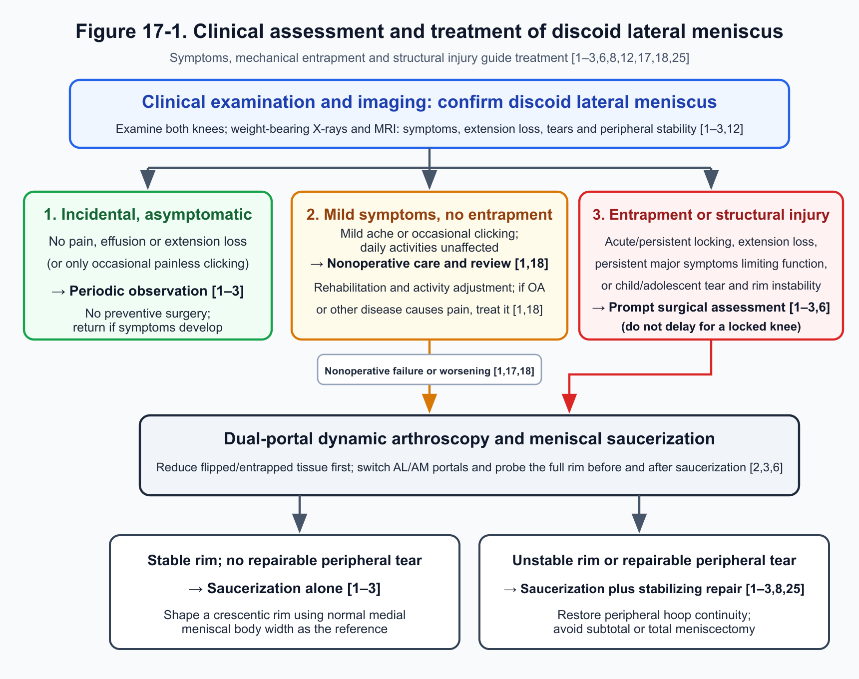
Figure 17-1. Clinical evaluation and treatment selection pathway for discoid lateral meniscus (based on references [1–3,6,8,12,17,18,25]).
For asymptomatic discoid lateral meniscus (for example, found incidentally during trauma evaluation or contralateral knee examination), the Chinese experts consensus (evidence level E) and mainstream international reviews are clear: prophylactic resection or saucerization is not recommended; periodic observation is preferred[1–3]. Intact, peripherally stable discoid menisci are mostly asymptomatic long term, and the knee cartilage and bony structure have already adapted mechanically; there is no evidence that prophylactic saucerization lowers the long-term tear rate, whereas resecting discoid meniscal tissue increases lateral compartment contact stress and induces valgus alignment shift[1,3,18]. (The Chinese experts consensus, in its evidence-level-E recommendation, also mentions physical therapy and avoidance of overload sports; these are expert protective suggestions, not a requirement to intervene medically in every asymptomatic incidental finding[1].) For patients with mild symptoms that do not affect daily life or general sports, without locking or extension deficit, and with normal lower-limb alignment, nonoperative treatment (rehabilitation and activity modification) with periodic follow-up is likewise preferred; if severe pain is driven mainly by concomitant osteoarthritis or rheumatoid arthritis, treatment should target the primary joint disease[1,18].
Arthroscopic surgical indications must integrate symptom severity, mechanical signs, and patient age[1–3,6,18]:
- Patients with clear mechanical entrapment signs such as acute or persistent locking or restricted passive extension (flexion contracture) should undergo timely arthroscopic exploration, reduction, and saucerization–repair, without mechanically waiting 3–6 months of nonoperative treatment[1–3,6].
- Patients with persistent lateral joint-line pain, painful snapping, or recurrent effusion that severely affects daily life and sports, or who lack acute locking but fail 3–6 months of structured nonoperative treatment with worsening symptoms, have clear indications for arthroscopic surgery (evidence level B2)[1,17,18].
- For children and adolescents with imaging or examination findings suggesting concomitant meniscal tear or peripheral instability, the Chinese experts consensus recommends early arthroscopic repair even when current symptoms are mild, to restore peripheral stability and reduce secondary cartilage wear (evidence level B2)[1].
When arguing for early surgery in children (p. 922, reference 134), the Chinese experts consensus cites Logan et al. (2021) as reporting that "the failure rate of conservative treatment for pediatric discoid meniscus lesions is as high as 39%"; this wording is detached from the denominator and population scope of that figure in the original study[1,11]. Logan et al. (2021, pp. 496–498 and Fig. 1), before including 401 symptomatic children (470 knees) from Boston Children's Hospital, had already excluded 13 asymptomatic incidental findings; of the 470 symptomatic knees, 387 went directly to surgery at first presentation, and only 83 knees (18%) initially underwent nonoperative observation, physical therapy, and/or bracing, of which 32 (32/83, 39%) converted to surgery at a median of 7.9 months because of persistent symptoms. Of the 51 knees that did not convert, 15 were lost to follow-up; of the 36 with last recorded status, 10 (27.8%; printed as 29% in the original) still had persistent symptoms (the same passage also prints physical therapy 30/83 [36.1%] as 37% and bracing 10/83 [12.0%] as 8%, and the Discussion on page 500 miswrites the 32 conversions as 33)[11]. Thus 39% is only the conversion-to-surgery rate among the 83 knees initially treated nonoperatively in that single-center symptomatic cohort (32/83); it cannot be applied directly to all symptomatic children or extended to asymptomatic discoid meniscus, nor can the remaining 61% who did not convert to surgery at that hospital be equated with cure by conservative treatment[1,11].
4. Arthroscopic Exploration, Saucerization, and Peripheral Stabilization Repair
Dual-Portal Dynamic Assessment and Intraoperative Definition of Peripheral Stability
The first step in discoid lateral meniscus surgery is thorough exploration and reduction; central tissue must not be resected before the peripheral attachment status is clear[2,6]. After standard anterolateral and anteromedial portals are established, if the lateral compartment is tight, the anteromedial portal may be placed slightly more proximal and medial than usual so that instruments can clear the tibial spines[6]. For menisci flipped and entrapped in the notch, popliteal hiatus, or posterior compartment, anatomic reduction with a probe must come first; if tension is high and a probe alone cannot maintain reduction, one peripheral traction suture may be placed before assessing the resection margin, to avoid injuring peripheral circumferential fibers in a twisted, folded state[2,6].
Intraoperative probing must cover the full circumference of the anterior horn, body, and posterior horn. Viewing from the anterolateral portal with the probe in the anteromedial portal facilitates assessment of plateau coverage and body thickness and forward traction of the posterior horn to test posterior-half stability, but viewing from the anterolateral portal alone leaves a blind zone for the anterior horn and easily misses anterior-half peripheral detachment[6,10]. Following the PRiSM standardized arthroscopic probing steps (Beck et al., 2022, pp. e1348–e1350), the arthroscope should routinely be switched to the anteromedial portal and the probe placed through the anterolateral portal; under direct vision the anterior horn of the lateral meniscus is pushed posteriorly and toward the joint center, with dynamic observation through flexion and extension, to confirm anterior-half peripheral instability[6]. Hypertrophic central discoid tissue may brace and mask a lax periphery before resection, and cutting into the central substance may also expose occult horizontal cleavage or peripheral longitudinal tears; probe stability assessment must therefore be completed both before and after saucerization[3,6].
Two common operational definitions of "peripheral instability" appear in the literature: the PRiSM scheme (Beck et al. 2022; Lee et al. 2022) defines it as absent, attenuated, or vertically split meniscocapsular attachment allowing the probe to push or pull the meniscus past the midpoint of the convexity of the lateral femoral condyle (the weight-bearing peak)[6,7]; Perkins et al. (2021, p. 24) define it as a tear within 3 mm of the meniscocapsular junction that the probe can displace toward the center of the tibial plateau (while noting that an intact synovial bridge on the surface may mask deep peripheral detachment)[8]. Operative records should specify the site and extent of probe-driven displacement.
Meniscal Saucerization and Suture Repair of Substance Cleavage and Peripheral Detachment
Meniscal saucerization (also called reshaping, meniscoplasty, or central partial meniscectomy) removes excess central discoid tissue in one piece or piecemeal, finely trims the free edge with basket forceps, curved scissors, or ophthalmic scissors, and smooths with a shaver so that the residual lateral meniscus recovers a crescentic contour and a wedge-shaped cross section that tapers medially. The normal medial meniscal body width in the same knee is often used as a visual anatomic reference[1–3].
After the central substance is opened and preliminary saucerization completed, myxoid degeneration or horizontal cleavage within the discoid meniscus is often exposed, leaving the residual peripheral rim as separated superior and inferior leaflets[3,6]. To avoid substantial loss of rim thickness from resecting a single leaflet, Campbell et al. (2023, pp. 157–158) advocate thorough clearance of loose interleaflet myxoid tissue with a curette or shaver, freshening the cleft with a rasp and performing trephination within the cleft to promote bleeding, then placing nonabsorbable vertical mattress (haybale) sutures every 4–5 mm to compress the superior and inferior leaflets and restore overall rim thickness[3]. (Conflict-of-interest note: the corresponding author of the 2023 Campbell et al. review, J.L. Pace, discloses consulting for Arthrex and JRF Ortho [p. 160]; named brand all-inside suture devices and advocacy of early meniscal transplantation in the asymptomatic period in that paper reflect author preference and lack supporting controlled-trial evidence[3].)
After saucerization, whenever probing confirms peripheral instability or a repairable tear in the red–red or red–white zone, combined peripheral stabilization suture repair (saucerization and repair) should be performed arthroscopically[1–3,8]. Suture technique is chosen by the site of detachment: anterior horn to anterior body peripheral detachment is usually addressed with outside-in suture or all-inside suture through a far-medial accessory portal; body to posterolateral corner, with inside-out double-needle cannula suture or all-inside suture; posterior horn and popliteal hiatus region peripheral detachment can be fixed with vertical mattress sutures using a suture hook or all-inside device viewed from anteromedial and placed through anterolateral or posterolateral portals, typically at 3–4 mm intervals[2,3,8]. After suturing, the probe must again be used segment by segment to confirm that the rim no longer passes the midpoint of the lateral femoral condyle convexity. For isolated meniscal repair with intact cartilage and ligaments, some surgeons perform marrow venting in a non–weight-bearing area of the notch to promote biologic healing, but its independent benefit in discoid meniscus has not been confirmed in controlled trials[3,8].
When interpreting multicenter cohort regression data on suture technique and peripheral instability prognosis, the methodologic limits of observational association must not be overlooked. Ohlsen et al. (2026, pp. 4–5), in a multivariable analysis of the North American 867-knee cohort, reported that open meniscal repair was associated with reoperation (OR = 19.50, 95% CI 1.52–250.71, P = 0.022), whereas baseline peripheral instability showed no statistically significant association with postoperative complications or reoperation[19]. The authors note in the Discussion that only 7 patients from 2 centers in the whole cohort underwent open repair (the preceding Sheasley et al. 2024 Table 1 on p. 2760 combines "open or transtibial repair" as 10 knees, a different statistical scope); the very small sample and selection bias toward complex tears produce an extremely wide confidence interval, so this OR cannot be treated as causal proof comparing surgical techniques. Likewise, because every detected instance of peripheral instability in that cohort received intraoperative repair, there is no "peripheral instability without repair" control group; failure to find a significant association between baseline peripheral instability and adverse events neither proves event rates equivalent to stable knees nor constitutes controlled evidence of treatment efficacy[9,19].
Controversies Regarding Subtotal Meniscectomy, Total Meniscectomy, and Meniscal Allograft Transplantation
Subtotal meniscectomy and total meniscectomy remove most of the weight-bearing body and peripheral circumferential collagen fibers, severely weakening hoop-stress transmission and accelerating lateral compartment degeneration[1,18,25,27]. Subtotal meniscectomy usually means residual peripheral rim width <3 mm; Cho et al. 2024 defined subtotal meniscectomy as preserving 2–3 mm and total meniscectomy as <2 mm, and grouped residual rim <6 mm as a sacrificed-rim group. The Chinese experts consensus (evidence levels B2 and C) states clearly that in both children/adolescents and adults, whenever peripheral instability or tear is amenable to suture and residual tissue quality is acceptable, saucerization combined with suture repair should be preferred and subtotal or total meniscectomy avoided[1].
Subtotal or total meniscectomy is reserved as salvage only when the meniscus shows severe global myxoid degeneration, a pulverized complex tear, or severe posterolateral corner deficiency that cannot be reduced with a probe and has completely lost conditions for suture fixation, or in elderly, low-demand patients with severe degeneration who have failed conservative treatment (evidence level C)[1,2,18]. For young patients with prior subtotal or total meniscectomy, persistent lateral compartment pain, and high activity demand, lateral meniscal allograft transplantation (MAT) may be considered; if progressive genu valgum has already developed, concomitant or staged distal femoral osteotomy is needed to unload the lateral compartment[1–3]. Whether outcomes of MAT using a normal-morphology donor after prior total meniscectomy for DLM are inferior to those after prior nondiscoid lateral meniscus (NDLM) resection differs substantially between two original controlled studies and cannot be summarized with the blanket statements of reviews[2,3,30,31]:
-
In a small controlled study, patient subjective scores showed no significant between-group differences, but range of motion was slightly smaller in the discoid group (Yoon et al., 2014): From 68 lateral MATs, Yoon et al.[30] excluded 27 combined procedures, 3 lost to follow-up, and 2 revisions, then compared 16 prior DLM with 20 prior NDLM treated by keyhole bone-bridge MAT (limited to patients with normal lower-limb alignment and mostly age <45 years; follow-up at least 2 years). Both groups improved significantly from preoperative to postoperative VAS, IKDC subjective, Lysholm, and Tegner scores; at final follow-up (Table 3, p. 728), the two groups did not differ significantly in VAS (2.6 vs 2.8), IKDC subjective (70.0 vs 69.4), Lysholm (78.8 vs 77.7), or Tegner (4.5 vs 4.2), whereas postoperative knee range of motion was smaller in the discoid group (132.8° vs 140.0°, P<0.05). In the subgroup of only 17 patients (9 vs 8) who completed postoperative MRI (body text p. 727 and Table 4 p. 729), absolute graft extrusion (2.0 mm vs 2.4 mm), relative extrusion percentage (22.8% vs 22.7%), and graft MRI signal grade likewise showed no significant between-group differences[30]. Given the small sample of 16 versus 20 (MRI subset only 9 vs 8), nonsignificant results do not constitute proof of equivalence[30]. Yoon et al. also give mutually inconsistent mean follow-up times in three places: the Abstract on page 724 states a whole-cohort mean of 32 months; the Results body on pages 726–727 states 37 months for the discoid group and 35 months for the nondiscoid group; Table 1 on page 727 prints 38 months for discoid and 41 months for nondiscoid—so no single mean can be treated as verified follow-up duration (additionally, a cross-column Results sentence on page 727 wrongly describes postoperative increases in IKDC, Lysholm, and Tegner together with VAS as having "decreased significantly"; the whole-cohort preoperative IKDC mean of 55.7 on the same page is inconsistent with the weighted value 55.1 from the two groups' 54.7 and 55.5 in Table 3; the MRI follow-up range of 6 months to 6 years stated in the body on page 727 [minimum 7.3 months in the discoid group] disagrees with the case-by-case range of 6–60 months overall and 11–32 months in the discoid group listed in Table 4 on page 729)[30].
-
A larger controlled study showed worse pain, subjective function scores, and extrusion in the discoid group, without significant difference in activity level (Ren et al., 2021): Ren et al.[31] included 106 unilateral lateral MAT patients (DLM group 59, NDLM group 47; age 18–50 years, no marked osteoarthritis at surgery; mean follow-up 31.3 months [range 24–36 months]). The paper reports that both groups improved significantly from preoperative to postoperative VAS, IKDC subjective, and Lysholm scores, whereas Tegner score showed no significant change (p. 4149); at final follow-up (Tables 2 and 3, p. 4151), the discoid group was significantly worse than the nondiscoid group in IKDC subjective (78.2 vs 82.6), Lysholm (81.0 vs 85.7), and VAS pain (3.1 vs 2.4), and coronal MRI absolute graft extrusion (3.7 mm vs 3.1 mm) and relative extrusion percentage (37.4% vs 30.5%) were also significantly greater, but postoperative Tegner medians were 6 in both groups without significant difference[31]. Campbell et al. (2023, p. 159) review summarizes Yoon and Ren together as showing that all clinical outcomes (including Tegner) were worse in the discoid group—clearly too broad[3,30,31]. Multivariable stepwise regression in Ren et al. (Table 4, p. 4151) showed that femoral condylar prominence ratio (condylar cut-off sign) and squaring of the lateral femoral condyle were significantly associated with final Lysholm score; this is an observational statistical association and cannot directly prove that osseous morphologic abnormality is the causal mechanism of between-group outcome differences[31]. In reporting and interpreting results, Ren et al. in the Discussion on page 4152 borrow minimal clinically important difference (MCID) and patient acceptable symptom state (PASS) thresholds from cartilage microfracture and ACL reconstruction and assert that the mean IKDC difference between groups in this study "certainly [has] no clinical significance"; individual improvement thresholds or state-attainment lines from other procedures cannot serve directly as equivalence margins for between-group mean comparisons in MAT (nor, conversely, can one assert that the difference has definite clinical significance). Furthermore, in Table 1 on page 4148 the discoid group's left/right laterality "12/18" sums to only 30 (not 59), and the nondiscoid group's "13/21" sums to only 34 (not 47); on the same page, Fig. 1's nondiscoid bone-bridge trough (BBT 23) and modified bone-plug technique (MBPT 24) counts are exactly reversed relative to the Methods body on page 4147 and Table 1 on page 4148 (24 and 23)[31] (Fig. 1 on page 4148 excludes 21 cases while listing three reasons totaling 11+12+1=24 without explaining overlap; the body on page 4149 contains the self-contradictory annotation "significantly better (n.s.)"; Table 4 on page 4151 prints P as 0.000; and the first paragraph of the Discussion on the same page retains a "Query" typesetting remnant)[31].
5. Anatomic and Imaging Basis for Residual Rim Width After Saucerization
In meniscal saucerization, "how many millimeters of peripheral rim should be preserved" is the most contested practical question. Traditional experience often recommends preserving 6–8 mm intraoperatively (the Chinese experts consensus summarizes the literature as usually preserving 6–10 mm, evidence level B2)[1,2]; some recent authors call for preserving 10 mm or more[3,20,23]. To understand these numbers accurately, four categories of "width" that differ completely in measurement site and time point must be distinguished (Fig. 17-2): the 45° oblique radial width of normal cadaveric specimens at the popliteal hiatus; the preserved width compared with a probe at a stated site during arthroscopy; coronal midbody MRI width at 1 week postoperatively ("postoperative month 0"); and coronal midbody MRI width at a stated long-term follow-up time point[17,20–23].
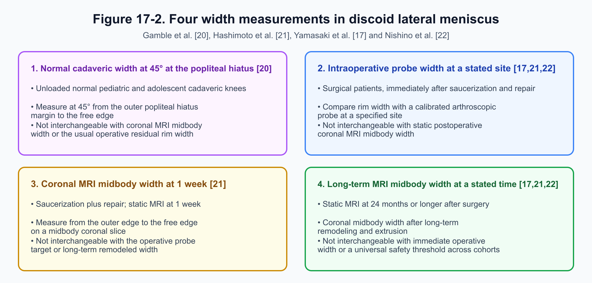
Figure 17-2. Four categories of width measurement and time points for discoid lateral meniscus (compiled from Gamble et al. 2022, Yamasaki et al. 2017, Hashimoto et al. 2026, and Nishino et al. 2022[17,20–22]; anatomic planes and follow-up times differ, so millimeter values are not interchangeable).
Measurement Differences Between Normal Anatomic Width and Early Postoperative MRI Width
Anatomic support for allowing a somewhat larger residual rim in adolescents comes from Gamble et al. (2022, pp. 138–141), who measured pediatric and adolescent cadaveric knees[20]. Measuring the radial width of the normal lateral meniscus from the outer edge of the popliteal hiatus to the free edge along a plane 45° to the coronal plane, they found that this oblique width already reached 10 mm or more by age 8, 15 mm or more in adolescence, and a mean of about 16 mm after skeletal maturity, suggesting that mechanically applying 6–8 mm in older children and adolescents may undershoot normal lateral meniscal volume[20]. Gamble et al. explicitly note, however, that unloaded cadaveric width in the 45° oblique plane at the popliteal hiatus (sample sizes and standard deviations by age band are not provided) cannot be equated with clinical coronal midbody MRI width, still less with a clinically prognosis-validated saucerization resection threshold[20]. When Hashimoto et al. (2026, Discussion p. 10) restate Gamble et al. (2022), they miswrite the conclusion as "a residual rim width of 6–8 mm is inadequate for children under 8 years," then in the next sentence write at least 10 mm at age 8 and at least 15 mm in adolescents; Gamble et al. tested and rejected the null hypothesis for "age 8 years and older (≥ 8 years)"—citations must not perpetuate the age-direction slip in Hashimoto's Discussion[20,21].
Preserved width measured with an arthroscopic probe differs substantially from midbody width on early postoperative coronal MRI. Hashimoto et al. (2026; enrollment 2017–2023; 72 patients, 77 knees with complete discoid lateral meniscus treated by saucerization plus repair; follow-up ≥2 years) had three surgeons uniformly target 8–10 mm with the probe, then grouped patients by coronal midbody MRI width at 1 week postoperatively ("postoperative month 0") into a large group (>13 mm, 21 knees), intermediate-width group (9–13 mm, 30 knees), and small group (<9 mm, 26 knees)[21]. Table 3 on page 6 shows that mean probe-measured preserved widths among the three groups were very close—9.8 mm, 9.2 mm, and 8.8 mm—yet mean midbody widths on 1-week coronal MRI reached 14.6 mm, 10.9 mm, and 7.5 mm, respectively, with only weak correlation between intraoperative probe width and 1-week MRI width (r = 0.332, P = 0.030)[21]. This confirms that the millimeter reading compared visually at the free edge with a probe is not the same scale as coronal MRI cross-sectional width after suture fixation (the original paper did not directly measure the mechanism of the difference, so it cannot simply be attributed to tissue edema or weight bearing alone)[21].
Relationship of Postoperative Rim Narrowing and Extrusion to Adverse Clinical Events
After discoid lateral meniscus saucerization, the residual peripheral rim commonly narrows over time under joint load and displaces laterally toward the capsule (extrusion):
-
Long-term postoperative MRI rim narrowing (<5.0 mm) and extrusion (>2.0 mm): Yamasaki et al. from Osaka City University (2017; enrollment 2007–2013; 40 patients, 45 adolescent saucerization knees; mean follow-up 39.6 months) set probe-preserved width at 6–8 mm intraoperatively (p. 571); by final follow-up, mean coronal MRI midbody width had narrowed to 4.6 mm (saucerization alone 5.2 mm; saucerization plus repair 4.1 mm). Using concurrent radiographic Tapper–Hoover grade 2 degeneration (7/45 knees) as the endpoint, the ROC cutoff for final MRI rim width was 5.0 mm (sensitivity 88%, specificity 68%)[17]. Nishino et al. from the same institution (2022; enrollment 2009–2017, overlapping enrollment period with the Yamasaki cohort; 39 patients, 48 skeletally immature complete discoid meniscus knees; likewise probe-preserved 6–8 mm) found mean midbody MRI width of 5.5 mm at 24 months postoperatively, with midbody extrusion increasing from 0.8 mm preoperatively to 1.6 mm; using lateral compartment cartilage degeneration on 24-month MRI (WORMS ≥ grade 3, 16 knees) as the endpoint, the ROC cutoff for postoperative extrusion was 2.0 mm (OR = 6.18 per 1 mm increase)[22]. These two studies show that 5.0 mm is a cross-sectional boundary value at which rim narrowing on long-term MRI after intraoperative preservation of 6–8 mm coexists with concurrent degeneration; it must not be read backwards as "preserving 5 mm at surgery is safe"[17,22]. Data reporting in the two papers also contains direct contradictions: Yamasaki et al. write final MRI rim width as "mean 4.6 mm (range 3.8–6.0 mm)" in the Abstract on page 570 but "4.6 ± 1.8 mm" in the Results body on page 573; if all 45 knees lie within a full range of 2.2 mm (3.8–6.0 mm), an SD of 1.8 mm is mathematically impossible—range and SD cannot both stand; when listing the negative correlation between rim width and relative extrusion percentage (Table 3 whole cohort r = −0.416, P = 0.004—i.e., the narrower the rim, the greater the relative extrusion), the Results body on page 573 wrongly calls it a "significant positive correlation"[17]. Nishino et al. wrongly call the positive correlation between postoperative extrusion and change in WORMS degeneration score (r = 0.418—greater extrusion associated with worse degeneration) a "significant negative correlation" in the body on page 5, and miswrite the 16 knees reaching the degeneration endpoint as "16 patients" in the Results body on page 4 (Table 1 on page 3 also adds 23 males and 25 females as 48 knees rather than 39 persons)[22].
-
Rapid narrowing and complication association when 1-week MRI midbody width is excessive (>13 mm): In the Hashimoto et al. (2026) cohort, the large group with 1-week coronal MRI midbody width >13 mm (mean 14.6 mm) narrowed by 55.7% to 6.4 mm at 24 months—nearly the same residual width as the intermediate-width group (9–13 mm; 6.2 mm at 24 months) (40.3% of all knees narrowed by more than 50% at 24 months; the ROC cutoff of 1-week MRI width for predicting >50% narrowing at 24 months was 11.5 mm)[21]. Table 2 on page 6 shows that the postoperative complication rate (retear, reoperation, and similar) in the large group (>13 mm) (6/21 knees, 28.6%) exceeded that in the intermediate-width group (1/30 knees, 3.3%) and the small group (2/26 knees, 7.7%)[21]. The authors speculate that if saucerization leaves excessive central discoid tissue with disordered collagen and myxoid degeneration, that region may still undergo accelerated degenerative resorption or retear under shear[21]. This is mechanistic discussion of observational grouping by 1-week MRI and cannot be converted into an operative threshold. Multiple statistical parameters in Hashimoto et al. also conflict between tables and text: for the same endpoint—"postoperative month 0 MRI midbody width predicting >50% width narrowing at 24 months"—the Abstract on page 1 reports multivariable OR = 1.38 (P = 0.006), whereas the Results body and Table 4 on page 9 report OR = 1.47 (95% CI 1.13–1.90, P = 0.003); AUC for the same ROC curve is 0.728 in the body and inside Fig. 4 on page 9 but 0.737 in the Fig. 4 legend on page 10; the overall P for three-group complication comparison appears as three different values—Table 2 p. 6 (P = 0.040), Results body p. 4 (P = 0.029), and Abstract p. 1 (P = 0.026) (the Discussion on page 10 also misprints the small-group complication rate 2/26 [7.7%] as 8.3%; page 4 miswrites 34 males and 43 females totaling 77 knees as numbers of patients; and Lysholm MCID calculated by the half-SD distribution method cannot prove clinical equivalence among the three groups)[21].
-
Methodologic limits of a single-arm long-term cohort with intraoperative probe preservation ≥10 mm: Aksoy et al. (2025) reviewed 53 patients (57 knees; mean age at surgery 27.6 years [6–65 years]; mean follow-up 12.1 years; ≤18 years 31 knees, >18 years 26 knees; only 6 knees combined with repair) after "limited saucerization (intraoperative probe preservation of at least 10 mm of rim)" and reported 4 reoperations at a mean of 5.7 years (2 arthroscopic reoperations, 2 total knee arthroplasties for osteoarthritis), with 5- and 10-year reoperation-free survival of 96.5% and 90.6%[23]. This is a single-arm case series without a control group preserving 6–8 mm or 8–10 mm intraoperatively, so it cannot prove that probe preservation ≥10 mm is superior to 8–10 mm; after completing survival analysis, the authors also directly excluded the 4 reoperated (treatment-failure) patients from the main analyses of subsequent clinical function scores and radiographic degeneration (main analysis left with only 53 knees: 29 pediatric, 24 adult; see p. 818 and Table 5), systematically overestimating final Lysholm scores and underestimating degeneration[23]. Even among the 53 knees after excluding failures, lateral joint-space width still narrowed significantly from preoperative values in both pediatric and adult groups, and final Kellgren–Lawrence (K–L) grade progressed significantly in adults (among 24 knees, grade 2 in 10 and grade 3 in 5); given that baseline cartilage degeneration was already heavier in adults than in children, one cannot infer that prophylactic early surgery in asymptomatic children would prevent adult degeneration[23].
Surgical Decision Principles for Residual Rim Width
Synthesizing available anatomic and clinical evidence, the literature values of 6–8 mm, 8–10 mm, and ≥10 mm each have specific measurement planes and study contexts; no prospective controlled trial has yet established a single millimeter cutoff applicable to all ages and body sizes[1,2,17,20,21,23]. When deciding the saucerization resection margin intraoperatively, a single fixed millimeter number should not be applied mechanically; three principles should be followed. First, thoroughly remove redundant central tissue that is free, laminated, or severely myxoid; if superior and inferior leaflets are separated at the periphery, debride and compress them with encircling sutures. Second, use the normal medial meniscal body width in the same knee as a visual anatomic reference and fashion a smooth crescentic contour with a medially tapering wedge slope (intraoperative probe reference often falls between 6 and 10 mm; in older adolescents with good substance quality, more healthy tissue may be preserved in light of anatomic width). Third, after saucerization, dual-portal probe dynamic testing of residual peripheral rim stability is mandatory, with secure suture fixation of any peripheral detachment[1–3,6,20,21].
6. Valgus Shift of Lower-Limb Alignment and Lateral Compartment Degeneration
The hypertrophic body of a discoid lateral meniscus occupies the lateral compartment and shares axial load; patients often present preoperatively with mild varus or reduced physiologic valgus[1,18]. After saucerization, subtotal meniscectomy, or total meniscectomy reduces lateral meniscal thickness and coverage, lateral compartment contact stress rises and the lower-limb mechanical axis tends to shift into valgus—especially in adults and when the resection is large (residual rim <6 mm)[1,3,18].
Cho et al. (2024) compared standing full-length radiographic alignment and lateral compartment degeneration progression among three groups of adults aged 20–65 years (mean 41.7 years; mean follow-up 47.6 months): a discoid lateral meniscus surgery group of 134 knees (all failures of 3–6 months of nonoperative treatment or worsening symptoms; including a preserved-rim >6 mm group of 108 knees and a sacrificed-rim <6 mm group of 26 knees), a discoid lateral meniscus nonoperative group of 56 knees (mild, tolerable symptoms), and a nondiscoid lateral meniscus tear surgery group of 64 knees[18]. Results showed postoperative valgus shift of the mechanical hip–knee–ankle angle (mHKA) in the discoid surgery group, and lateral compartment osteoarthritis progression (K–L grade advance to 3–4) in 54/134 knees (40.3%) in the discoid surgery group, higher than 11/64 (17.2%) in the nondiscoid surgery group and 4/56 (7.1%) in the mild discoid nonoperative group; within the discoid surgery group, patients older than 40 years (56.3% vs ≤40 years 16.7%) and those with sacrificed rim <6 mm (69.2% vs preserved rim >6 mm 33.3%) had significantly higher rates of lateral degeneration progression[18].
When interpreting Cho et al. (2024), indication confounding is the primary issue. The nonoperative group (56 knees) comprised patients with mild symptoms and relatively stable structure, whereas the surgery group (134 knees) comprised patients who had failed conservative treatment, had tears, or had severe symptoms; the degeneration difference of 40.3% versus 7.1% jointly reflects baseline disease severity and tissue removed at surgery and cannot be read, apart from severity, as "avoiding surgery for symptomatic adult discoid meniscus tear prevents degeneration"[18].
Whole-cohort means and age-subgroup means within that paper's tables contradict each other. In Table 2 on page 1227, mean mHKA change for all 134 discoid surgery knees is +3.23° ± 1.85°, which matches the weighted mean 3.23° from the preserved-rim group of 108 knees (2.86° ± 1.74°) and the sacrificed-rim group of 26 knees (4.79° ± 1.44°) in the lower half of Table 3 on pages 1228–1229; yet in the upper half of the same Table 3, when the same 134 knees are split by age into >40 years (80 knees) and ≤40 years (54 knees), mean mHKA change is printed as 4.68° ± 1.98° (one place in the body on page 1228 prints the SD as 1.85) and 2.96° ± 2.00°, which weight to a whole-cohort mean change of 3.99°. Weighted preoperative and postoperative mHKA means of the two age groups likewise cannot recover the Table 2 whole-cohort means, whereas weighted mPTA, mLDFA, and JLCA on the same table match Table 2. The Table 2 whole-cohort mean (+3.23°) and Table 3 age-subgroup means (4.68° and 2.96°) cannot both stand after weighted checking (likely a reporting or typesetting error; the original calculation process cannot be located)[18]; clinically one may credit only the direction of postoperative valgus shift and the trend toward greater shift with residual rim <6 mm, not treat the above angle means as verified exact values (the Abstract on page 1223 and Discussion on page 1228 also miswrite the nonoperative group's degeneration progression of 4/56 [7.1%] from Table 2 as 5.3%)[18].
Regarding postoperative lower-limb alignment change, Wen et al. (2026) completed a systematic review and meta-analysis of 11 studies (including Cho et al. 2024)[24]. Across all between-group comparisons in Table 2 on page 6, only the weighted mean difference in postoperative change in medial proximal tibial angle (mPTA) between discoid and nondiscoid groups reached statistical significance (WMD = −0.20°, 95% CI −0.35° to −0.05°, 2 studies; the original paper broadly calls this negative difference a "smaller improvement effect"); pooled alignment results across resection extents showed no statistically significant differences, but between-study heterogeneity was extremely high (I² often exceeding 70%) and confidence intervals were wide, so one cannot conclude that total meniscectomy and rim-preserving saucerization affect lower-limb alignment equally[24]. When reporting the single-group change in tibiofemoral angle (TFA) after meniscal saucerization, Wen et al. print 0.24° (95% CI 1.85°–2.63°) in the Results body on page 5—the point estimate 0.24° lying entirely outside its own confidence interval—whereas the same cell in Table 2 on page 6 prints 2.24° (95% CI 1.85°–2.63°); body and table conflict directly, and neither value can be selected alone from this meta-analysis as a reliable pooled summary of TFA change after saucerization[24].
7. Mid- to Long-Term Outcomes, Retear, and Complications
Effect of Follow-up Duration on Clinical Outcomes and Postoperative Complications
After discoid lateral meniscus saucerization with or without suture repair, short-term function scores at 2–4 years postoperatively (e.g., Lysholm, Pedi-IKDC) mostly improve substantially; when follow-up extends to 5–20 years, rates of rim retear, reoperation, osteochondritis dissecans (OCD) of the lateral femoral condyle in adolescents, and lateral compartment degeneration are relatively high[4,5,8,9,11,25,26]. Interpreting related cohorts requires strict distinction among follow-up duration, crude incidence versus Kaplan–Meier cumulative incidence, patient-number versus knee-number denominators, and overlapping same-center cohorts:
-
Intermediate outcomes after peripheral repair with rim preservation, and observational comparison of osteochondritis dissecans: In a pediatric–adolescent cohort dedicated to saucerization plus suture repair for peripheral instability, Perkins et al. (2021) followed 30 patients (32 knees, mean age 12 years) for a mean of 54 months; 3/32 knees (9.4%) underwent further meniscal surgery; in 1 of these, repeat arthroscopy confirmed complete healing of the original peripheral repair with only a new central horizontal tear treated by partial resection—showing that reoperation does not equal failure of the index peripheral suture to heal (among 28 patients completing scale follow-up, final Pedi-IKDC averaged 96, and 89% returned to preoperative sport level)[8]. Shimmyo et al. (2025) compared a later institutional period (2011–2018) of pediatric–adolescent saucerization plus suture repair (27 knees, 21 patients, mean follow-up 50.6 months) with an earlier period (2005–2011) of subtotal meniscectomy (rim <3 mm; 22 knees, 20 patients, mean follow-up 62.3 months) as a historical control: both groups showed significant narrowing of the operated-side lateral joint space from preoperative values; retear occurred in 5/27 knees (19%) after saucerization plus repair and in 0/22 after subtotal meniscectomy (because subtotal meniscectomy leaves a stump under 3 mm, almost no meniscal substance remains in which retear can occur—this cannot be taken to show that subtotal meniscectomy is superior to suture repair)[25]. Conversely, 6 knees in the subtotal meniscectomy group developed new OCD of the lateral femoral condyle, all requiring osteochondral surgical intervention, whereas only 1 knee in the saucerization-plus-repair group did so and healed with nonoperative treatment; these are differences in observed incidence and severity between historical cohorts from different eras, which differ in surgical era, indications, follow-up duration, and technique, and cannot be treated as confounding-free causal risk facts verified by controlled study[25]. Additionally, Shimmyo et al. note in a footnote to Table 4 on page 6 that 1 patient in each group with preoperative OCD was excluded when calculating postoperative new OCD (denominators after exclusion should be 26 knees for saucerization plus repair and 21 for subtotal meniscectomy), yet the "6 (27%)" printed for the subtotal meniscectomy group in Table 4 does not match 6/21 (28.6%) and corresponds instead to the pre-exclusion 6/22 (27.3%); as for the "1 (4%)" printed for saucerization plus repair, both 1/26 (3.8%) and 1/27 (3.7%) round to 4%, so one cannot conclude from "1 (4%)" that the repair group also used a wrong denominator, nor reverse-engineer from the printed percentage a test result (P = 0.036) whose exact calculation process is not disclosed[25].
-
Reoperation rates in multicenter and long-term follow-up cohorts, and conflicts in original reporting:
-
North American 9-center SCORE homologous cohort (Sheasley et al. 2024; Ohlsen et al. 2026): Ohlsen et al. state explicitly on page 2 that their analysis population is entirely homologous with Sheasley et al.—the same 784 patients (867 knees; mean age at diagnosis 12 years; mean follow-up 22.6 months); the two papers must never be added as independent cohorts[9,19]. Within a mean follow-up under 2 years, 105/784 patients (13%) and 110/867 knees (12.7%) underwent a total of 135 reoperations; in Ohlsen et al.'s two-level multivariable regression, female sex and complex tear both significantly raised risks of postoperative complications and reoperation, whereas horizontal tear was significantly associated only with raised reoperation risk (association with overall complications did not reach statistical significance)[19]. Meanwhile, Sheasley et al. (2024) state in the Results body on page 2760 that the whole cohort had 175 complication events involving 139 knees (134 patients; 116 knees with 1 complication and 23 with >1, 116 + 23 = 139), yet Table 1 lower on the same page lists total complications as 98 knees (11%; 75 with 1 and 23 with >1, 75 + 23 = 98; the same table also prints saucerization 837/867 [96.5%] as 98%); the subsequent same-cohort paper Ohlsen et al. (2026) directly enters 139/867 knees (16.0%) into the regression model without issuing an erratum for the 98 knees in the 2024 Table 1[9,19].
-
Boston Children's Hospital cohort (Logan et al. 2021; Lins et al. 2021; same institution and corresponding author; potential overlap for cases operated before the end of 2002): Among 379 surgically treated knees with postoperative follow-up, Logan et al. recorded reoperation in 66/379 knees (17%, involving 63 patients) (median time to first reoperation 19.6 months; meniscal retear in 62/66); among the 313 knees without reoperation, another 8 had retear, 1 a parameniscal cyst, and 51 persistent pain or mechanical symptoms; adding the 66 reoperations to the 60 without reoperation but still abnormal or symptomatic at last follow-up, the authors report a composite adverse outcome of 126/379 knees (33%). Citations must distinguish the true reoperation rate of 17% from the composite adverse outcome rate of 33%[11] (the body on page 498 also writes peripheral instability as "52% [116 knees]" while Table 2 and the Discussion give 164/316 [52%]; miswrites the denominator for complex tear 44/163 [27%] as 419 knees; mislabels the interquartile range 9.2–34.9 months after 19.6 months as the full range; miswrites 126/379 knees as patients; prints 470 knees as 471 on page 499; and on pages 498–499 writes a median interval of 1.2 years for 7 second reoperations while printing an interquartile range of 1.8–2.1 years[11]). Lins et al. assessed by questionnaire 25 patients (30 knees) operated before the end of 2002 with at least 15 years of follow-up (response rate 26%; mean follow-up 19.5 years) and found that 11/25 patients (44%, patient-number denominator) had undergone at least one subsequent ipsilateral operation, and that 13/25 (52%) had final Lysholm scores in the fair or poor range (Table 3 labels the IKDC score range as 0–87 without stating whether these are raw scores; Marx scale upper limits of 10 versus 16 also disagree between Abstract and Table 3)[5].
-
French pediatric rim-preserving saucerization cohort (Bonny Obro et al., 2025): Among 47 responding children (56 knees; mean age at surgery 8.6 years; all rim-preserving saucerization with or without suture), mean follow-up was 10.5 years; mean Lysholm rose from 72.9 to 90.5; postoperative OCD of the lateral femoral condyle occurred in 2 (both improved with activity restriction); the Abstract on page 295 and body on page 297 state that "10 patients (17.9%)" underwent arthroscopic revision at a mean of 2.7 years postoperatively, but 10/47 responding patients is 21.3% and 10/56 knees equals 17.9%—citations should state 10 cases, 47 persons, and 56 knees together[26].
-
Olmsted County, USA, population cohort (Sabbag et al., 2019): Following 59 surgically treated symptomatic discoid lateral meniscus patients (ages 4–66 years; mean 25.7 years; mean follow-up 5.6 years), Kaplan–Meier survival analysis estimated an 8-year cumulative retear rate of 59% (retear-free survival 93% at 2 years, 71% at 5 years, 41% at 8 years) and an 8-year cumulative reoperation rate of 39% (reoperation-free survival 61%); Cox proportional-hazards regression showed that younger age at surgery and open physes in this cohort were associated with higher postoperative retear risk (the Methods and Table 3 on page 4 do not describe a multivariable adjustment model; Table 1 also prints open physes 16/59 [27.1%] as 17%, and operative-category classifications disagree between body and Table 1)[4]. Sabbag et al. also define clearly in the Methods on page 2 that the endpoint "progression to symptomatic lateral compartment osteoarthritis" requires both persistent knee symptoms and final radiograph Kellgren–Lawrence grade of 2 or higher (K–L ≥ 2); yet the Results paragraph on page 3 states that among the 18 patients counted toward this "symptomatic osteoarthritis" endpoint, only 11 (61%) reached K–L ≥ 2 on the final radiograph. Without individual survival data one cannot confirm how the remaining 7 were coded in the statistical analysis, but the results report is directly incompatible with the prespecified methodologic definition, so the paper's "8-year cumulative incidence of symptomatic osteoarthritis of 50%" cannot be cited as a verified incidence of K–L ≥ 2 osteoarthritis[4].
Serious Errors in Systematic Reviews Comparing Surgical Techniques and Extracting Data
Three recent systematic reviews (Hartman et al. 2026; Vivekanantha et al. 2025; Mohan et al. 2024) contain multiple primary-data extraction errors and denominator confusions that affect their main conclusions when summarizing surgical outcomes of discoid lateral meniscus; clinicians must not cite their pooled percentages directly to judge the relative merits of different techniques[27–29]:
Hartman et al. (2026, Arthroscopy)—complication and reoperation rates by technique group are wholly unusable because of severe extraction errors: Table 4 on page 1893 splits Lins et al. (2021, Table 2 p. e812—whole cohort only 30 knees: saucerization alone 20, saucerization plus repair 10)[5] into a saucerization-alone group of 30 knees (with 8 reoperations) and a saucerization-plus-repair group of 20 knees (with 3 reoperations), inventing an expansion to 50 knees; it also wrongly assigns 20 knees from Yamasaki et al. (2017, p. 571—all 45 knees were rim-preserving saucerization, with no subtotal or total meniscectomy)[17] (together with the whole cohort's 5 reoperations and 1 OCD) to the third group (total and subtotal meniscectomy); and for the third group's 4 studies totaling 157 knees, 32 complications (actual 32/157 = 20.4%), and 8 reoperations (actual 8/157 = 5.1%), the Results body on page 1891 prints complication rate 14.2% and reoperation rate 8.0%, and in the same paragraph first calls the saucerization-plus-repair group (6.8%) as having the "highest" complication rate, then in the next sentence writes the third group's complication rate as 14.2%[27] (additionally, page 1887 writes inclusion of 24 articles / 1,225 persons while quality appraisal and Discussion write 25 articles / 1,236 persons; the follow-up range 1.2–10.1 years omits Lins's 19.5 years in Table 2; page 1888 prints 551/1,401 [39.3%] as 39.1% and 723/1,401 [51.6%] as 51.4%; and a Table 4 footnote expands osteochondritis OCD wrongly as "obsessive-compulsive disorder")[27].
Vivekanantha et al. (2025, Curr Rev Musculoskelet Med)—the body dilutes pooled incidence by using the full-library total knee count that includes studies not reporting the outcome; the Abstract mixes inconsistent denominators within the same sentence; and the body seriously misquotes the Logan cohort reoperation rate: in the Results body on page 523 the authors first divide the library-wide totals of 209 retears and 290 reoperations by the full 52-study sample of 4,784 knees, reporting overall retear 4.4% and reoperation 6.0%, and only then note that only 3,190 knees' studies reported retear (209/3,190 = 6.6%) and only 3,415 knees' studies reported reoperation (290/3,415 = 8.5%). In the Abstract on page 513, under the premise "Amongst patients with reported values," retear is listed as 6.6% (209) against the 3,190 knees that reported that outcome, yet reoperation is listed as 6.0% (290) against the full-library 4,784 knees rather than the 8.5% calculated from 3,415. Using a full-library denominator that includes 1,594 and 1,369 knees from studies not reporting the outcome directly dilutes pooled percentages and underestimates the postoperative complication burden[28]. The review also states on pages 530–532 that Logan et al. (2021) reported a reoperation rate of 33% among surgical patients, whereas the true postoperative reoperation rate in the Logan cohort is 17% (66/379 knees); 33% (126/379 knees) includes 60 knees without reoperation but still symptomatic at last follow-up; and Table 4 on page 531 further divides Logan's 62 retears and 66 reoperations by all 470 knees (including 51 nonsurgical and 40 lost to follow-up) to obtain 13.2% and 14.0%, omitting the additional 8 retears in the non-reoperated group (Table 1 on page 521 also mislabels the median first-reoperation interval of 19.6 [9.2–34.9] months as whole-cohort follow-up time; the weighted mean postoperative Tegner of 7.3 and VAS of 3.2 reported on page 523 both fall outside the ranges of the listed study means, 4.3–5.0 and 0.6–1.2)[11,28].
Mohan et al. (2024) pooled 12 articles with at least 5 years of follow-up (444 knees) and support that rim-preserving saucerization (with or without repair), compared with subtotal or total meniscectomy, reduces long-term lateral compartment degeneration; because radiographic degeneration grading differs across primary studies, and because that paper lists saucerization, reshaping, and meniscoplasty as different technique categories (page 621 also misprints central partial resection 198/444 [44.6%] as 43.9%), one cannot infer absence of lateral compartment degeneration progression from favorable subjective function scores alone[29].
8. Postoperative Rehabilitation Protection and Long-term Follow-up Requirements
Differentiated Rehabilitation Principles for Saucerization Alone Versus Combined Repair
Postoperative rehabilitation after discoid lateral meniscus surgery depends mainly on whether the meniscus was suture-repaired intraoperatively. No randomized controlled trial comparing rehabilitation protocols exists (evidence level C), but center protocols all distinguish early protection intensity by whether a suture–repair interface is present:
- Meniscal saucerization alone (and subtotal or total meniscectomy): There is no suture interface requiring protection; isometric quadriceps contraction, straight-leg raises, and range-of-motion exercises may begin the day after surgery, with early progression to full weight bearing as tolerated; once effusion resolves and quadriceps strength and neuromuscular control recover, jogging and routine sports may be resumed gradually[1,17,22,25,26].
- Meniscal saucerization combined with suture repair: The residual meniscus is thicker and the suture line bears substantial shear; early postoperatively, brace or cast limitation of knee flexion and delayed full weight bearing are mandatory, avoiding early deep squatting and other high-shear maneuvers (for example, Kim et al.[2] prohibit kneeling and deep squatting for 6 months postoperatively, whereas Perkins et al.[8] resume sports after 4 months)[1,2,8,17,21,22,26]. Progression speed must be individualized by suture site and tension, patient age and adherence, and clinical markers such as resolution of effusion, full recovery of extension, absence of joint-line tenderness, and recovery of strength. Representative rehabilitation protocols from the literature are listed in Table 17-1 (the rehabilitation text of Yamasaki et al. 2017[17] p. 572 is identical to that of Nishino et al. 2022[22] from the same institution; Bonny Obro et al. 2025[26] use 4–6 weeks of long-leg cast immobilization with non–weight bearing for suture patients under 10 years). Clinically, one coherent protocol should be chosen and adjusted as clinical milestones are met; weight-bearing and return-to-sport time points from different centers must not be mechanically spliced together.
Table 17-1. Comparison of representative postoperative rehabilitation protocols for discoid lateral meniscus
| Protocol source | Applicable procedure and population | Brace immobilization and ROM management | Weight-bearing progression | Return to sport (RTS) timing and special management |
|---|---|---|---|---|
| Chinese experts consensus (2023)[1] | Saucerization alone (or subtotal/total meniscectomy) | Knee brace immobilization not recommended; knee motion and isometric strengthening from postoperative day 1 | Full weight bearing by postoperative week 2; continuous active motion from week 4 to restore proprioception, stability, and strength promptly | Normal motor function by postoperative weeks 6–8 (normal motor; original does not specify months for return to specific sports) |
| Chinese experts consensus (2023)[1] | Saucerization combined with suture repair | Isometric strengthening from postoperative day 1; knee brace limiting ROM for the first 6 weeks; ROM unrestricted from week 7 | Full-weight-bearing rehabilitation and proprioceptive training at postoperative weeks 4–6 (original does not detail weight bearing in the first 3 weeks) | Gradual return to normal motor function at postoperative weeks 12–16 (normal motor; original does not specify months for return to specific sports) |
| Kim et al. (2020)[2] | Saucerization combined with suture repair | Progressive angle increase under brace protection from postoperative day 2; 120° flexion by postoperative week 8 | Crutch non–weight bearing for 4 weeks, then partial weight bearing for 4 weeks | Running allowed at 3 months; kneeling and deep squatting prohibited for 6 months; return to sport after confirming strength and neuromuscular coordination at 5–6 months (high-impact sports advised with caution) |
| Perkins et al. (2021)[8] | Pediatric–adolescent saucerization combined with peripheral repair | Knee immobilizer brace for 6 weeks; physical therapy started postoperative days 1–3; ROM limited to 0°–90° for the first 4 weeks, then advanced as tolerated | Crutch partial weight bearing for the first 2 weeks, then progression to full weight bearing under brace protection as tolerated | Sports usually resumed after 4 months postoperatively |
| Nishino et al. (2022)[22] | Saucerization alone | Knee ROM exercises allowed immediately postoperatively | Weight bearing allowed immediately postoperatively | Jogging allowed at 2 months; return to prior sports at 3 months |
| Nishino et al. (2022)[22] | Saucerization combined with suture stabilization | Brace immobilization for 1 week postoperatively, then ROM limited to 0°–90° for 3 weeks | After the above 4 weeks, followed by 6 weeks of protected weight bearing (protected weightbearing for 6 weeks; original does not separately state weight-bearing status during the first 4 weeks) | Jogging allowed at 3 months; return to prior sports at 6 months |
| Hashimoto et al. (2026)[21] | Saucerization combined with suture repair | Extension brace immobilization for 1 week postoperatively, then ROM limited to 0°–90° for 3 weeks | After 1 week of brace immobilization, nonweightbearing for 3 weeks, then partial weightbearing for 6 weeks | Jogging allowed at 3 months; return to prior sports at 6 months (deferred by 1 month if follow-up MRI shows marked effusion, cartilage injury, or bone marrow edema) |
Standardized Operative Records and Long-term Follow-up Monitoring
Because postoperative rim narrowing, extrusion, lower-limb valgus shift, adolescent lateral femoral condyle OCD, and adult lateral compartment osteoarthritis can all progress slowly over years to more than a decade after discoid lateral meniscus surgery, clinical follow-up should not end when the patient returns to sport at about six months[1,4,5,18,23,25].
To ensure long-term follow-up and cross-center data are verifiable, the index operative record should completely document five measured items: first, lateral tibial plateau coverage (complete or incomplete) and thickness of the body and free edge; second, anterior horn, body, posterior horn, and popliteal hiatus region peripheral stability assessed dynamically with a probe through dual anterolateral and anteromedial portals (specifying whether, before and after saucerization, the probe could displace the meniscus past the midpoint of the lateral femoral condyle convexity); third, tear type within the residual peripheral rim (horizontal cleavage, vertical longitudinal, radial, or complex) and the zone involved; fourth, specific sites, techniques, and number of sutures used for repair; fifth, residual rim width in millimeters measured with a probe scale at the end of saucerization and the exact measurement site[6–8,12,20].
Postoperative imaging follow-up should combine standing knee radiographs (standing full-length lower-limb anteroposterior radiographs at re-evaluation for older adolescents and adults) with MRI[1,18]. Per the 2026 SSR standardized nomenclature consensus (Nguyen et al., pp. 1714, 1718), postoperative MRI reports should avoid the ambiguous term "residual tear" and instead distinguish expected signal change after saucerization and repair from signal suggesting new retear; when measuring meniscal extrusion, state the lateral compartment, coronal reference slice, and millimeters; when recording postoperative MRI rim width, the exact time from surgery must be stated (e.g., 1 week or 2 years postoperatively)[12,21,22].
References
- [1]
Yang S, Zhang S, Li R, et al. Chinese experts consensus and practice guideline on discoid lateral meniscus. Orthop Surg. 2023;15(4):915-929. doi:10.1111/os.13687. PMID:36878896.
- [2]
Kim JH, Ahn JH, Kim JH, Wang JH. Discoid lateral meniscus: importance, diagnosis, and treatment. J Exp Orthop. 2020;7(1):81. doi:10.1186/s40634-020-00294-y. PMID:33044686.
- [3]
Campbell AL, Pace JL, Mandelbaum BR. Discoid lateral meniscus. Curr Rev Musculoskelet Med. 2023;16(4):154-161. doi:10.1007/s12178-023-09824-4. PMID:36920747.
- [4]
Sabbag OD, Hevesi M, Sanders TL, et al. High rate of recurrent meniscal tear and lateral compartment osteoarthritis in patients treated for symptomatic lateral discoid meniscus: a population-based study. Orthop J Sports Med. 2019;7(7):2325967119856284. doi:10.1177/2325967119856284. PMID:31367646.
- [5]
Lins LAB, Feroe AG, Yang B, et al. Long-term minimum 15-year follow-up after lateral discoid meniscus rim preservation surgery in children and adolescents. J Pediatr Orthop. 2021;41(9):e810-e815. doi:10.1097/BPO.0000000000001903. PMID:34411050.
- [6]
Beck JJ, Schlechter J, Schmale G, Haus B, Lee J. Comprehensive arthroscopic characterization of discoid meniscus tears and instability using the PRiSM discoid meniscus classification. Arthrosc Tech. 2022;11(7):e1347-e1352. doi:10.1016/j.eats.2022.03.018. PMID:35936837.
- [7]
Lee RJ, Nepple JJ, Schmale GA, et al. Reliability of a new arthroscopic discoid lateral meniscus classification system: a multicenter video analysis. Am J Sports Med. 2022;50(5):1245-1253. doi:10.1177/03635465221076857. PMID:35234542.
- [8]
Perkins CA, Busch MT, Christino MA, Willimon SC. Saucerization and repair of discoid lateral menisci with peripheral rim instability: intermediate-term outcomes in children and adolescents. J Pediatr Orthop. 2021;41(1):23-27. doi:10.1097/BPO.0000000000001695. PMID:33044260.
- [9]
Sheasley JA, Kirby JC, Niu EL, et al. Characteristics and outcomes of operatively treated discoid lateral meniscus in pediatric and young adult patients: a multicenter study. Am J Sports Med. 2024;52(11):2758-2763. doi:10.1177/03635465241272393. PMID:39222125.
- [10]
Henick SM, Samuel Z, Charla JN, et al. Anterior peripheral rim instability is prevalent in young, skeletally immature patients with discoid lateral meniscus and has favorable postoperative outcomes. Arthrosc Sports Med Rehabil. 2025;7(3):101119. doi:10.1016/j.asmr.2025.101119. PMID:40692925.
- [11]
Logan CA, Tepolt FA, Kocher SD, et al. Symptomatic discoid meniscus in children and adolescents: a review of 470 cases. J Pediatr Orthop. 2021;41(8):496-501. doi:10.1097/BPO.0000000000001907. PMID:34397782.
- [12]
Nguyen JC, Gorbachova T, Flores DV, et al. Standardized MRI nomenclature for knee meniscal lesions: SSR consensus panel report. Skeletal Radiol. 2026;55(8):1711-1732. doi:10.1007/s00256-026-05281-5. PMID:42283751.
- [13]
You M, Li J, Zhang X, Chen G, Li J. Diagnostic efficiency of MRI in child and adolescent lateral discoid meniscus. Orthop Surg. 2025;17(3):900-908. doi:10.1111/os.14357. PMID:39924921.
- [14]
Niu EL, Milewski MD, Finlayson CJ, et al. Reliability of MRI interpretation of discoid lateral meniscus: a multicenter study. Orthop J Sports Med. 2023;11(5):23259671231174475. doi:10.1177/23259671231174475. PMID:37275780.
- [15]
Sun XX, Zhou W, Zuo SP, et al. MRI morphologic indices for evaluating injury of the lateral discoid meniscus in children. Zhongguo Zuzhi Gongcheng Yanjiu. 2017;21(7):1104-1109. doi:10.3969/j.issn.2095-4344.2017.07.022.
- [16]
Amiri S, Mirahmadi A, Parvandi A, et al. Excellent accuracy of magnetic resonance imaging for diagnosis of discoid meniscus tears: a systematic review and meta-analysis. J Exp Orthop. 2024;11(3):e12051. doi:10.1002/jeo2.12051. PMID:38899047.
- [17]
Yamasaki S, Hashimoto Y, Takigami J, et al. Risk factors associated with knee joint degeneration after arthroscopic reshaping for juvenile discoid lateral meniscus. Am J Sports Med. 2017;45(3):570-577. doi:10.1177/0363546516668623. PMID:28272930.
- [18]
Cho JH, Nam HS, Park SY, Ho JPY, Lee YS. Arthroscopic meniscal repair and meniscectomy for adult discoid lateral meniscus results in progression to valgus alignment and lateral compartment degeneration compared with nonoperative treatment and nondiscoid lateral meniscus. Arthroscopy. 2024;40(4):1223-1233. doi:10.1016/j.arthro.2023.08.085. PMID:37717929.
- [19]
Ohlsen S, Son M, Howard W, et al. Risk factors for complication and reoperation in the treatment of discoid lateral meniscus in children and adolescents: a multicenter study. Orthop J Sports Med. 2026;14(2):23259671251411397. doi:10.1177/23259671251411397. PMID:41727782.
- [20]
Gamble JG, Abdalla AB, Meadows MG, et al. Radial width of the lateral meniscus at the popliteal hiatus: relevance to saucerization of discoid lateral menisci. Am J Sports Med. 2022;50(1):138-141. doi:10.1177/03635465211056661. PMID:34780308.
- [21]
Hashimoto Y, Aibara N, Yamasaki S, Nishida Y, Tomihara T, Terai H. Residual midbody meniscal widths >13 mm immediately after surgery decreased faster and had greater complication rates in discoid lateral menisci treated with saucerization and repair. Arthroscopy. 2026. doi:10.1002/arj.70378. PMID:42383583.
- [22]
Nishino K, Hashimoto Y, Iida K, Nishida Y, Yamasaki S, Nakamura H. Association of postoperative lateral meniscal extrusion with cartilage degeneration on magnetic resonance imaging after discoid lateral meniscus reshaping surgery. Orthop J Sports Med. 2022;10(5):23259671221091997. doi:10.1177/23259671221091997. PMID:35528992.
- [23]
Aksoy T, Goymen IM, Huri G, Turhan E, Kocher MS, Atay OA. Midterm to long-term follow-up after limited saucerization of a discoid lateral meniscus: radiological and functional outcomes with age-dependent variations. Am J Sports Med. 2025;53(4):817-825. doi:10.1177/03635465241313137. PMID:39881482.
- [24]
Wen C, Hua Q, Qian W, Su J, Lei M. Effects of arthroscopic meniscectomy or meniscoplasty of discoid meniscus on lower limb axial alignment: a systematic review and meta-analysis. BMC Musculoskelet Disord. 2026;27(1):258. doi:10.1186/s12891-026-09611-5. PMID:41723375.
- [25]
Shimmyo A, Onishi S, Kanto R, et al. Combined meniscal saucerization and repair versus subtotal meniscectomy for symptomatic discoid lateral meniscal tears in children and adolescents. Orthop J Sports Med. 2025;13(2):23259671241311918. doi:10.1177/23259671241311918. PMID:39931636.
- [26]
Bonny Obro R, Rassi J, Flumian C, Sales de Gauzy J, Accadbled F. Arthroscopic management of discoid lateral meniscus in children: 5-year minimum follow-up clinical outcome. J Child Orthop. 2025;19(4):295-300. doi:10.1177/18632521251325030. PMID:40692963.
- [27]
Hartman H, Lessiohadi N, Saharan S, et al. Outcomes of saucerization with or without repair for symptomatic discoid lateral meniscus in pediatric patients: a systematic review. Arthroscopy. 2026;42(9):1882-1899. doi:10.1002/arj.70391. PMID:42478088.
- [28]
Vivekanantha P, Thomas R, Kaplan G, et al. Surgical management of the discoid lateral meniscus: a systematic review of outcomes. Curr Rev Musculoskelet Med. 2025;18(11):513-535. doi:10.1007/s12178-025-09980-9. PMID:40388072.
- [29]
Mohan K, Bharti A, Kumar M, et al. Long term surgical and radiological outcome of discoid lateral meniscus: an updated systematic review. Indian J Orthop. 2024;58(6):619-636. doi:10.1007/s43465-024-01137-z. PMID:38812876.
- [30]
Yoon KH, Lee SH, Park SY, Jung GY, Chung KY. Meniscus allograft transplantation for discoid lateral meniscus: clinical comparison between discoid lateral meniscus and nondiscoid lateral meniscus. Arthroscopy. 2014;30(6):724-730. doi:10.1016/j.arthro.2014.02.017. PMID:24768466.
- [31]
Ren S, Zhou R, Zhang X, et al. Anatomical knee variables result in worse outcomes of lateral meniscal allograft transplantation with discoid lateral menisci than with nondiscoid lateral menisci. Knee Surg Sports Traumatol Arthrosc. 2021;29(12):4146-4153. doi:10.1007/s00167-021-06509-8. PMID:33660054.